6UY4 | pdb_00006uy4

Crystal structure of dihydroorotate dehydrogenase from Schistosoma mansoni


Experimental Data Snapshot

  • Method: X-RAY DIFFRACTION
  • Resolution: 2.80 Å
  • R-Value Free: 
    0.246 (Depositor), 0.245 (DCC) 
  • R-Value Work: 
    0.216 (Depositor), 0.216 (DCC) 
  • R-Value Observed: 
    0.217 (Depositor) 

Starting Model: experimental
View more details

wwPDB Validation   3D Report Full Report


Ligand Structure Quality Assessment 


This is version 1.2 of the entry. See complete history


Literature

Structural basis for the function and inhibition of dihydroorotate dehydrogenase from Schistosoma mansoni.

de Mori, R.M.Aleixo, M.A.A.Zapata, L.C.C.Calil, F.A.Emery, F.S.Nonato, M.C.

(2021) FEBS J 288: 930-944

  • DOI: https://doi.org/10.1111/febs.15367
  • Primary Citation Related Structures: 
    6UY4

  • PubMed Abstract: 

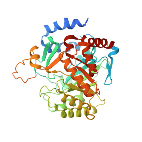
    Schistosomiasis is a serious public health problem, prevalent in tropical and subtropical areas, especially in poor communities without access to safe drinking water and adequate sanitation. Transmission has been reported in 78 countries, and its control depends on a single drug, praziquantel, which has been used over the past 30 years. Our work is focused on exploiting target-based drug discovery strategies to develop new therapeutics to treat schistosomiasis. In particular, we are interested in evaluating the enzyme dihydroorotate dehydrogenase (DHODH) as a drug target. DHODH is a flavoenzyme that catalyzes the stereospecific oxidation of (S)-dihydroorotate (DHO) to orotate during the fourth and only redox step of the de novo pyrimidine nucleotide biosynthetic pathway. Previously, we identified atovaquone, used in the treatment of malaria, and its analogues, as potent and selective inhibitors against Schistosoma mansoni DHODH (SmDHODH). In the present article, we report the first crystal structure of SmDHODH in complex with the atovaquone analogue inhibitor 2-((4-fluorophenyl)amino)-3-hydroxynaphthalene-1,4-dione (QLA). We discuss three major findings: (a) the open conformation of the active site loop and the unveiling of a novel transient druggable pocket for class 2 DHODHs; (b) the presence of a protuberant domain, only present in Schistosoma spp DHODHs, that was found to control and modulate the dynamics of the inhibitor binding site; (c) a detailed description of an unexpected binding mode for the atovaquone analogue to SmDHODH. Our findings contribute to the understanding of the catalytic mechanism performed by class 2 DHODHs and provide the molecular basis for structure-guided design of SmDHODH inhibitors. DATABASE: The structural data are available in Protein Data Bank (PDB) database under the accession code number 6UY4.


  • Organizational Affiliation
    • Laboratório de Cristalografia de Proteínas, Faculdade de Ciências Farmacêuticas de Ribeirão Preto, Universidade de São Paulo, Ribeirão Preto, Brazil.

Macromolecules
Find similar proteins by:  (by identity cutoff)  |  3D Structure
Entity ID: 1
MoleculeChains Sequence LengthOrganismDetailsImage
Dihydroorotate dehydrogenase379Schistosoma mansoniMutation(s): 0 
Gene Names: Smp_078730
EC: 1.3.98.1 (PDB Primary Data), 1.3.5.2 (UniProt)
UniProt
Find proteins for G4VFD7 (Schistosoma mansoni)
Explore G4VFD7 
Go to UniProtKB:  G4VFD7
Entity Groups  
Sequence Clusters30% Identity50% Identity70% Identity90% Identity95% Identity100% Identity
UniProt GroupG4VFD7
Sequence Annotations
Expand
  • Reference Sequence
Small Molecules
Ligands 3 Unique
IDChains Name / Formula / InChI Key2D Diagram3D Interactions
FMN
Query on FMN

Download Ideal Coordinates CCD File 
G [auth A]FLAVIN MONONUCLEOTIDE
C17 H21 N4 O9 P
FVTCRASFADXXNN-SCRDCRAPSA-N
QLA (Subject of Investigation/LOI)
Query on QLA

Download Ideal Coordinates CCD File 
B [auth A]2-[(4-fluorophenyl)amino]-3-hydroxynaphthalene-1,4-dione
C16 H10 F N O3
AVGHWZUEYVXFON-UHFFFAOYSA-N
GOL
Query on GOL

Download Ideal Coordinates CCD File 
C [auth A],
D [auth A],
E [auth A],
F [auth A]
GLYCEROL
C3 H8 O3
PEDCQBHIVMGVHV-UHFFFAOYSA-N
Binding Affinity Annotations 
IDSourceBinding Affinity
QLA BindingDB:  6UY4 IC50: 1000 (nM) from 1 assay(s)
Experimental Data & Validation

Experimental Data

  • Method: X-RAY DIFFRACTION
  • Resolution: 2.80 Å
  • R-Value Free:  0.246 (Depositor), 0.245 (DCC) 
  • R-Value Work:  0.216 (Depositor), 0.216 (DCC) 
  • R-Value Observed: 0.217 (Depositor) 
Space Group: I 4 3 2
Unit Cell:
Length ( Å )Angle ( ˚ )
a = 187.981α = 90
b = 187.981β = 90
c = 187.981γ = 90
Software Package:
Software NamePurpose
PHENIXrefinement
PDB_EXTRACTdata extraction
XDSdata reduction
Aimlessdata scaling
PHASERphasing

Structure Validation

View Full Validation Report


Ligand Structure Quality Assessment 


Entry History & Funding Information

Deposition Data


Funding OrganizationLocationGrant Number
Sao Paulo Research Foundation (FAPESP)Brazil2019/04395-6
Sao Paulo Research Foundation (FAPESP)Brazil2019/08376-6

Revision History  (Full details and data files)

  • Version 1.0: 2020-05-27
    Type: Initial release
  • Version 1.1: 2021-03-03
    Changes: Database references
  • Version 1.2: 2023-10-11
    Changes: Data collection, Database references, Refinement description