6UUM | pdb_00006uum

Crystal structure of antibody 438-B11 DSS mutant (Cys98A-Cys100aA)

  • Classification: IMMUNE SYSTEM
  • Organism(s): Homo sapiens
  • Expression System: Homo sapiens
  • Mutation(s): No 

  • Deposited: 2019-10-30 Released: 2020-09-23 
  • Deposition Author(s): Kumar, S., Wilson, I.A.
  • Funding Organization(s): National Institutes of Health/National Institute Of Allergy and Infectious Diseases (NIH/NIAID)

Experimental Data Snapshot

  • Method: X-RAY DIFFRACTION
  • Resolution: 2.10 Å
  • R-Value Free: 
    0.269 (Depositor), 0.269 (DCC) 
  • R-Value Work: 
    0.229 (Depositor), 0.230 (DCC) 
  • R-Value Observed: 
    0.230 (Depositor) 

Starting Model: experimental
View more details

wwPDB Validation 3D Report Full Report

Validation slider image for 6UUM

Ligand Structure Quality Assessment 


This is version 1.3 of the entry. See complete history

Literature

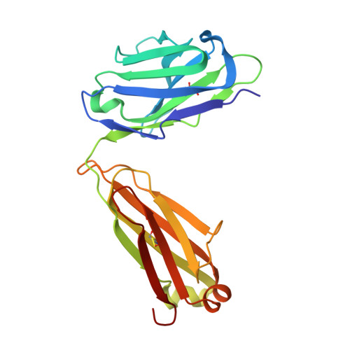
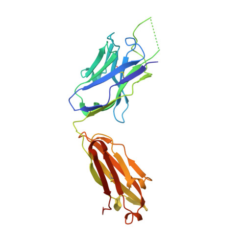
A V H 1-69 antibody lineage from an infected Chinese donor potently neutralizes HIV-1 by targeting the V3 glycan supersite.

Kumar, S.Ju, B.Shapero, B.Lin, X.Ren, L.Zhang, L.Li, D.Zhou, Z.Feng, Y.Sou, C.Mann, C.J.Hao, Y.Sarkar, A.Hou, J.Nunnally, C.Hong, K.Wang, S.Ge, X.Su, B.Landais, E.Sok, D.Zwick, M.B.He, L.Zhu, J.Wilson, I.A.Shao, Y.

(2020) Sci Adv 6

  • DOI: https://doi.org/10.1126/sciadv.abb1328
  • Primary Citation Related Structures: 
    6UUH, 6UUL, 6UUM

  • PubMed Abstract: 

    An oligomannose patch around the V3 base of HIV-1 envelope glycoprotein (Env) is recognized by multiple classes of broadly neutralizing antibodies (bNAbs). Here, we investigated the bNAb response to the V3 glycan supersite in an HIV-1-infected Chinese donor by Env-specific single B cell sorting, structural and functional studies, and longitudinal analysis of antibody and virus repertoires. Monoclonal antibodies 438-B11 and 438-D5 were isolated that potently neutralize HIV-1 with moderate breadth, are encoded by the V H 1-69 germline gene, and have a disulfide-linked long HCDR3 loop. Crystal structures of Env-bound and unbound antibodies revealed heavy chain-mediated recognition of the glycan supersite with a unique angle of approach and a critical role of the intra-HCDR3 disulfide. The mechanism of viral escape was examined via single-genome amplification/sequencing and glycan mutations around the N332 supersite. Our findings further emphasize the V3 glycan supersite as a prominent target for Env-based vaccine design.


  • Organizational Affiliation
    • Department of Integrative Structural and Computational Biology, The Scripps Research Institute, La Jolla, CA 92037, USA.

Macromolecule Content 

  • Total Structure Weight: 101.78 kDa 
  • Atom Count: 7,498 
  • Modeled Residue Count: 884 
  • Deposited Residue Count: 900 
  • Unique protein chains: 2

Macromolecules

Find similar proteins by:|  3D Structure
Entity ID: 1
MoleculeChains  Sequence LengthOrganismDetailsImage
B11 Fab Light ChainA [auth F],
C [auth A]
215Homo sapiensMutation(s): 0 
Find similar proteins by:|  3D Structure
Entity ID: 2
MoleculeChains  Sequence LengthOrganismDetailsImage
B11 DSS Fab Heavy ChainB [auth H],
D [auth B]
235Homo sapiensMutation(s): 0 

Small Molecules

Ligands 4 Unique
IDChains Name / Formula / InChI Key2D Diagram3D Interactions
NAG

Query on NAG


Download:Ideal Coordinates CCD File
AA [auth A],
G [auth F]
2-acetamido-2-deoxy-beta-D-glucopyranose
C8 H15 N O6
OVRNDRQMDRJTHS-FMDGEEDCSA-N
PG4
(Subject of Investigation/LOI)

Query on PG4


Download:Ideal Coordinates CCD File
BA [auth B]
CA [auth B]
DA [auth B]
E [auth F]
F
BA [auth B],
CA [auth B],
DA [auth B],
E [auth F],
F,
H [auth F],
Q [auth H],
R [auth H],
S [auth H],
T [auth H],
X [auth A],
Y [auth A],
Z [auth A]
TETRAETHYLENE GLYCOL
C8 H18 O5
UWHCKJMYHZGTIT-UHFFFAOYSA-N
SO4

Query on SO4


Download:Ideal Coordinates CCD File
EA [auth B]
FA [auth B]
GA [auth B]
HA [auth B]
I [auth F]
EA [auth B],
FA [auth B],
GA [auth B],
HA [auth B],
I [auth F],
J [auth F],
K [auth F],
L [auth F],
M [auth F],
N [auth F],
O [auth F],
U [auth H],
V [auth H],
W [auth H]
SULFATE ION
O4 S
QAOWNCQODCNURD-UHFFFAOYSA-L
ACT

Query on ACT


Download:Ideal Coordinates CCD File
IA [auth B],
P [auth F]
ACETATE ION
C2 H3 O2
QTBSBXVTEAMEQO-UHFFFAOYSA-M

Experimental Data & Validation

Experimental Data

  • Method: X-RAY DIFFRACTION
  • Resolution: 2.10 Å
  • R-Value Free:  0.269 (Depositor), 0.269 (DCC) 
  • R-Value Work:  0.229 (Depositor), 0.230 (DCC) 
  • R-Value Observed: 0.230 (Depositor) 
Space Group: P 1 21 1
Unit Cell:
Length ( Å )Angle ( ˚ )
a = 104.151α = 90
b = 66.197β = 117.959
c = 108.856γ = 90
Software Package:
Software NamePurpose
PHENIXrefinement
Cootmodel building
HKL-2000data reduction
SCALEPACKdata scaling
PHASERphasing

Structure Validation

View Full Validation Report


Ligand Structure Quality Assessment 


Entry History 

& Funding Information

Deposition Data


Funding OrganizationLocationGrant Number
National Institutes of Health/National Institute Of Allergy and Infectious Diseases (NIH/NIAID)United States--

Revision History  (Full details and data files)

  • Version 1.0: 2020-09-23
    Type: Initial release
  • Version 1.1: 2020-09-30
    Changes: Database references
  • Version 1.2: 2023-10-11
    Changes: Data collection, Database references, Refinement description
  • Version 1.3: 2024-11-20
    Changes: Structure summary