6URW | pdb_00006urw

Crystal structure of ricin A chain in complex with inhibitor 4-(2-thienylmethyl)benzoic acid


Experimental Data Snapshot

  • Method: X-RAY DIFFRACTION
  • Resolution: 2.40 Å
  • R-Value Free: 
    0.235 (Depositor), 0.235 (DCC) 
  • R-Value Work: 
    0.184 (Depositor), 0.190 (DCC) 
  • R-Value Observed: 
    0.187 (Depositor) 

Starting Model: experimental
View more details

wwPDB Validation 3D Report Full Report

Validation slider image for 6URW

Ligand Structure Quality Assessment 


This is version 1.2 of the entry. See complete history

Literature

Small Molecule Inhibitors Targeting the Interaction of Ricin Toxin A Subunit with Ribosomes.

Li, X.P.Harijan, R.K.Kahn, J.N.Schramm, V.L.Tumer, N.E.

(2020) ACS Infect Dis 6: 1894-1905

  • DOI: https://doi.org/10.1021/acsinfecdis.0c00127
  • Primary Citation Related Structures: 
    6URW, 6URX, 6URY

  • PubMed Abstract: 

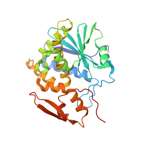
    Ricin toxin A subunit (RTA) removes an adenine from the universally conserved sarcin/ricin loop (SRL) on eukaryotic ribosomes, thereby inhibiting protein synthesis. No high affinity and selective small molecule therapeutic antidotes have been reported against ricin toxicity. RTA binds to the ribosomal P stalk to access the SRL. The interaction anchors RTA to the P protein C-termini at a well-defined hydrophobic pocket, which is on the opposite face relative to the active site. The RTA ribosome binding site has not been previously targeted by small molecule inhibitors. We used fragment screening with surface plasmon resonance to identify small molecular weight lead compounds that bind RTA and defined their interactions by crystallography. We identified five fragments, which bound RTA with mid-micromolar affinity. Three chemically distinct binding fragments were cocrystallized with RTA, and crystal structures were solved. Two fragments bound at the P stalk binding site, and the third bound to helix D, a motif distinct from the P stalk binding site. All fragments bound RTA remote from the catalytic site and caused little change in catalytic site geometry. Two fragments uniquely bound at the hydrophobic pocket with affinity sufficient to inhibit the catalytic activity on eukaryotic ribosomes in the low micromolar range. The binding mode of these inhibitors mimicked the interaction of the P stalk peptide, establishing that small molecule inhibitors can inhibit RTA binding to the ribosome with the potential for therapeutic intervention.


  • Organizational Affiliation
    • Department of Plant Biology, Rutgers, The State University of New Jersey, 59 Dudley Road, New Brunswick, New Jersey 08901, United States.

Macromolecule Content 

  • Total Structure Weight: 31.73 kDa 
  • Atom Count: 2,247 
  • Modeled Residue Count: 260 
  • Deposited Residue Count: 268 
  • Unique protein chains: 1

Macromolecules

Find similar proteins by:|  3D Structure
Entity ID: 1
MoleculeChains  Sequence LengthOrganismDetailsImage
Ricin268Ricinus communisMutation(s): 0 
EC: 3.2.2.22
UniProt
Find proteins for P02879 (Ricinus communis)
Explore P02879 
Go to UniProtKB:  P02879
Entity Groups
Sequence Clusters30% Identity50% Identity70% Identity90% Identity95% Identity100% Identity
UniProt GroupP02879
Sequence Annotations
Expand
Reference Sequence

Small Molecules

Ligands 3 Unique
IDChains Name / Formula / InChI Key2D Diagram3D Interactions
R7T
(Subject of Investigation/LOI)

Query on R7T


Download:Ideal Coordinates CCD File
Y [auth A]4-[(thiophen-2-yl)methyl]benzoic acid
C12 H10 O2 S
OAIBKSRZDOMJKZ-UHFFFAOYSA-N
DMS

Query on DMS


Download:Ideal Coordinates CCD File
X [auth A]DIMETHYL SULFOXIDE
C2 H6 O S
IAZDPXIOMUYVGZ-UHFFFAOYSA-N
EDO

Query on EDO


Download:Ideal Coordinates CCD File
B [auth A]
C [auth A]
D [auth A]
E [auth A]
F [auth A]
B [auth A],
C [auth A],
D [auth A],
E [auth A],
F [auth A],
G [auth A],
H [auth A],
I [auth A],
J [auth A],
K [auth A],
L [auth A],
M [auth A],
N [auth A],
O [auth A],
P [auth A],
Q [auth A],
R [auth A],
S [auth A],
T [auth A],
U [auth A],
V [auth A],
W [auth A]
1,2-ETHANEDIOL
C2 H6 O2
LYCAIKOWRPUZTN-UHFFFAOYSA-N

Experimental Data & Validation

Experimental Data

  • Method: X-RAY DIFFRACTION
  • Resolution: 2.40 Å
  • R-Value Free:  0.235 (Depositor), 0.235 (DCC) 
  • R-Value Work:  0.184 (Depositor), 0.190 (DCC) 
  • R-Value Observed: 0.187 (Depositor) 
Space Group: P 63 2 2
Unit Cell:
Length ( Å )Angle ( ˚ )
a = 168.615α = 90
b = 168.615β = 90
c = 54.657γ = 120
Software Package:
Software NamePurpose
REFMACrefinement
PDB_EXTRACTdata extraction
MOSFLMdata reduction
Aimlessdata scaling
PHASERphasing

Structure Validation

View Full Validation Report


Ligand Structure Quality Assessment 


Entry History 

& Funding Information

Deposition Data


Funding OrganizationLocationGrant Number
National Institutes of Health/National Institute Of Allergy and Infectious Diseases (NIH/NIAID)United States--

Revision History  (Full details and data files)

  • Version 1.0: 2020-06-17
    Type: Initial release
  • Version 1.1: 2020-07-22
    Changes: Database references
  • Version 1.2: 2023-10-11
    Changes: Data collection, Database references, Refinement description