6UN3

Crystal structure of Pseudomonas aeruginosa PBP3 in complex with ticarcillin

  • Classification: HYDROLASE
  • Organism(s): Pseudomonas aeruginosa
  • Expression System: Escherichia coli
  • Mutation(s): No 

  • Deposited: 2019-10-10 Released: 2019-10-30 
  • Deposition Author(s): Sacco, M., Chen, Y.
  • Funding Organization(s): National Institutes of Health/National Institute Of Allergy and Infectious Diseases (NIH/NIAID)

Experimental Data Snapshot

  • Method: X-RAY DIFFRACTION
  • Resolution: 1.90 Å
  • R-Value Free: 0.267 
  • R-Value Work: 0.201 
  • R-Value Observed: 0.204 

wwPDB Validation   3D Report Full Report


Ligand Structure Quality Assessment 


This is version 1.4 of the entry. See complete history


Literature

Influence of the alpha-Methoxy Group on the Reaction of Temocillin with Pseudomonas aeruginosa PBP3 and CTX-M-14 beta-Lactamase.

Sacco, M.D.Kroeck, K.G.Kemp, M.T.Zhang, X.Andrews, L.D.Chen, Y.

(2019) Antimicrob Agents Chemother 64

  • DOI: https://doi.org/10.1128/AAC.01473-19
  • Primary Citation of Related Structures:  
    6UN1, 6UN3, 6UNB

  • PubMed Abstract: 

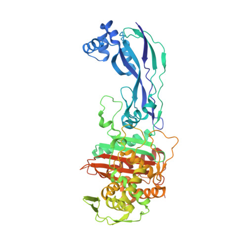
    The prevalence of multidrug-resistant Pseudomonas aeruginosa has led to the reexamination of older "forgotten" drugs, such as temocillin, for their ability to combat resistant microbes. Temocillin is the 6-α-methoxy analogue of ticarcillin, a carboxypenicillin with well-characterized antipseudomonal properties. The α-methoxy modification confers resistance to serine β-lactamases, yet temocillin is ineffective against P. aeruginosa growth. The origins of temocillin's inferior antibacterial properties against P. aeruginosa have remained relatively unexplored. Here, we analyze the reaction kinetics, protein stability, and binding conformations of temocillin and ticarcillin with penicillin-binding protein 3 (PBP3), an essential PBP in P. aeruginosa We show that the 6-α-methoxy group perturbs the stability of the PBP3 acyl-enzyme, which manifests in an elevated off-rate constant ( k off ) in biochemical assays comparing temocillin with ticarcillin. Complex crystal structures with PBP3 reveal similar binding modes of the two drugs but with important differences. Most notably, the 6-α-methoxy group disrupts a high-quality hydrogen bond with a conserved residue important for ligand binding while also being inserted into a crowded active site, possibly destabilizing the active site and enabling water molecule from bulk solvent to access and cleave the acyl-enzyme bond. This hypothesis is supported by the observation that the acyl-enzyme complex of temocillin has reduced thermal stability compared with ticarcillin. Furthermore, we explore temocillin's mechanism of β-lactamase inhibition with a high-resolution complex structure of CTX-M-14 class A serine β-lactamase. The results suggest that the α-methoxy group prevents hydrolysis by locking the compound into an unexpected conformation that impedes access of the catalytic water to the acyl-enzyme adduct.


  • Organizational Affiliation

    Department of Molecular Medicine, University of South Florida, Tampa, Florida, USA.


Macromolecules
Find similar proteins by:  (by identity cutoff)  |  3D Structure
Entity ID: 1
MoleculeChains Sequence LengthOrganismDetailsImage
Peptidoglycan D,D-transpeptidase FtsI533Pseudomonas aeruginosaMutation(s): 0 
Gene Names: 
EC: 3.4.16.4
UniProt
Find proteins for G3XD46 (Pseudomonas aeruginosa (strain ATCC 15692 / DSM 22644 / CIP 104116 / JCM 14847 / LMG 12228 / 1C / PRS 101 / PAO1))
Explore G3XD46 
Go to UniProtKB:  G3XD46
Entity Groups  
Sequence Clusters30% Identity50% Identity70% Identity90% Identity95% Identity100% Identity
UniProt GroupG3XD46
Sequence Annotations
Expand
  • Reference Sequence
Small Molecules
Ligands 3 Unique
IDChains Name / Formula / InChI Key2D Diagram3D Interactions
XT8 (Subject of Investigation/LOI)
Query on XT8

Download Ideal Coordinates CCD File 
C [auth A](2R,4S)-2-[(1R)-1-{[(2R)-2-carboxy-2-(thiophen-3-yl)acetyl]amino}-2-oxoethyl]-5,5-dimethyl-1,3-thiazolidine-4-carboxylic acid
C15 H18 N2 O6 S2
GQZADNJRZOHJDT-MWGHHZFTSA-N
GOL
Query on GOL

Download Ideal Coordinates CCD File 
B [auth A]GLYCEROL
C3 H8 O3
PEDCQBHIVMGVHV-UHFFFAOYSA-N
CA
Query on CA

Download Ideal Coordinates CCD File 
D [auth A]CALCIUM ION
Ca
BHPQYMZQTOCNFJ-UHFFFAOYSA-N
Experimental Data & Validation

Experimental Data

  • Method: X-RAY DIFFRACTION
  • Resolution: 1.90 Å
  • R-Value Free: 0.267 
  • R-Value Work: 0.201 
  • R-Value Observed: 0.204 
  • Space Group: P 21 21 21
Unit Cell:
Length ( Å )Angle ( ˚ )
a = 68.79α = 90
b = 83.26β = 90
c = 89.22γ = 90
Software Package:
Software NamePurpose
REFMACrefinement
SCALAdata scaling
PDB_EXTRACTdata extraction
MOSFLMdata reduction
MOLREPphasing

Structure Validation

View Full Validation Report


Ligand Structure Quality Assessment 


Entry History & Funding Information

Deposition Data


Funding OrganizationLocationGrant Number
National Institutes of Health/National Institute Of Allergy and Infectious Diseases (NIH/NIAID)United StatesR21 AI147654

Revision History  (Full details and data files)

  • Version 1.0: 2019-10-30
    Type: Initial release
  • Version 1.1: 2019-11-20
    Changes: Database references
  • Version 1.2: 2019-12-18
    Changes: Author supporting evidence
  • Version 1.3: 2020-01-01
    Changes: Database references
  • Version 1.4: 2023-10-11
    Changes: Data collection, Database references, Derived calculations, Refinement description