6UFP

Structure of proline utilization A with the FAD covalently modified by L-thiazolidine-2-carboxylate and three cysteines (Cys46, Cys470, Cys638) modified to S,S-(2-HYDROXYETHYL)THIOCYSTEINE


Experimental Data Snapshot

  • Method: X-RAY DIFFRACTION
  • Resolution: 1.74 Å
  • R-Value Free: 0.200 
  • R-Value Work: 0.168 
  • R-Value Observed: 0.169 

wwPDB Validation   3D Report Full Report


Ligand Structure Quality Assessment 


This is version 1.2 of the entry. See complete history


Literature

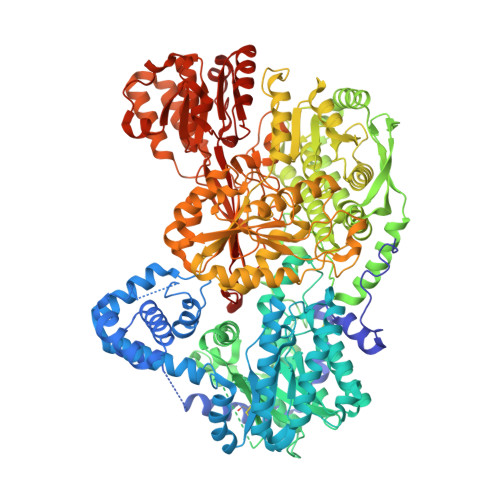
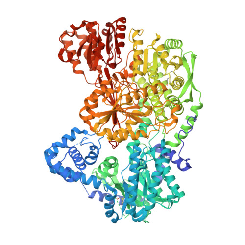
Covalent Modification of the Flavin in Proline Dehydrogenase by Thiazolidine-2-Carboxylate.

Campbell, A.C.Becker, D.F.Gates, K.S.Tanner, J.J.

(2020) ACS Chem Biol 15: 936-944

  • DOI: https://doi.org/10.1021/acschembio.9b00935
  • Primary Citation of Related Structures:  
    6UFP, 6VZ9

  • PubMed Abstract: 

    Proline dehydrogenase (PRODH) catalyzes the first step of proline catabolism, the FAD-dependent 2-electron oxidation of l-proline to Δ 1 -pyrroline-5-carboxylate. PRODH has emerged as a possible cancer therapy target, and thus the inhibition of PRODH is of interest. Here we show that the proline analogue thiazolidine-2-carboxylate (T2C) is a mechanism-based inactivator of PRODH. Structures of the bifunctional proline catabolic enzyme proline utilization A (PutA) determined from crystals grown in the presence of T2C feature strong electron density for a 5-membered ring species resembling l-T2C covalently bound to the N5 of the FAD in the PRODH domain. The modified FAD exhibits a large butterfly bend angle, indicating that the FAD is locked into the 2-electron reduced state. Reduction of the FAD is consistent with the crystals lacking the distinctive yellow color of the oxidized enzyme and stopped-flow kinetic data showing that T2C is a substrate for the PRODH domain of PutA. A mechanism is proposed in which PRODH catalyzes the oxidation of T2C at the C atom adjacent to the S atom of the thiazolidine ring (C5). Then, the N5 atom of the reduced FAD attacks the C5 of the oxidized T2C species, resulting in the covalent adduct observed in the crystal structure. To our knowledge, this is the first report of T2C inactivating (or inhibiting) PRODH or any other flavoenzyme. These results may inform the design of new mechanism-based inactivators of PRODH for use as chemical probes to study the roles of proline metabolism in cancer.


  • Organizational Affiliation

    Department of Biochemistry, University of Missouri, Columbia, Missouri 65211, United States.


Macromolecules
Find similar proteins by:  (by identity cutoff)  |  3D Structure
Entity ID: 1
MoleculeChains Sequence LengthOrganismDetailsImage
Bifunctional protein PutA1,235Sinorhizobium meliloti SM11Mutation(s): 0 
Gene Names: putASM11_chr0102
EC: 1.5.5.2 (PDB Primary Data), 1.2.1.88 (PDB Primary Data)
UniProt
Find proteins for F7X6I3 (Sinorhizobium meliloti (strain SM11))
Explore F7X6I3 
Go to UniProtKB:  F7X6I3
Entity Groups  
Sequence Clusters30% Identity50% Identity70% Identity90% Identity95% Identity100% Identity
UniProt GroupF7X6I3
Sequence Annotations
Expand
  • Reference Sequence
Find similar proteins by:  (by identity cutoff)  |  3D Structure
Entity ID: 2
MoleculeChains Sequence LengthOrganismDetailsImage
Bifunctional protein PutA1,235Sinorhizobium meliloti SM11Mutation(s): 0 
Gene Names: putASM11_chr0102
EC: 1.5.5.2 (PDB Primary Data), 1.2.1.88 (PDB Primary Data)
UniProt
Find proteins for F7X6I3 (Sinorhizobium meliloti (strain SM11))
Explore F7X6I3 
Go to UniProtKB:  F7X6I3
Entity Groups  
Sequence Clusters30% Identity50% Identity70% Identity90% Identity95% Identity100% Identity
UniProt GroupF7X6I3
Sequence Annotations
Expand
  • Reference Sequence
Small Molecules
Ligands 6 Unique
IDChains Name / Formula / InChI Key2D Diagram3D Interactions
FDA (Subject of Investigation/LOI)
Query on FDA

Download Ideal Coordinates CCD File 
C [auth A],
L [auth B]
DIHYDROFLAVINE-ADENINE DINUCLEOTIDE
C27 H35 N9 O15 P2
YPZRHBJKEMOYQH-UYBVJOGSSA-N
NAD (Subject of Investigation/LOI)
Query on NAD

Download Ideal Coordinates CCD File 
E [auth A],
N [auth B]
NICOTINAMIDE-ADENINE-DINUCLEOTIDE
C21 H27 N7 O14 P2
BAWFJGJZGIEFAR-NNYOXOHSSA-N
T2C (Subject of Investigation/LOI)
Query on T2C

Download Ideal Coordinates CCD File 
D [auth A],
M [auth B]
(2S)-1,3-thiazolidine-2-carboxylic acid
C4 H7 N O2 S
ULSZVNJBVJWEJE-VKHMYHEASA-N
PEG
Query on PEG

Download Ideal Coordinates CCD File 
F [auth A],
O [auth B]
DI(HYDROXYETHYL)ETHER
C4 H10 O3
MTHSVFCYNBDYFN-UHFFFAOYSA-N
SO4
Query on SO4

Download Ideal Coordinates CCD File 
G [auth A]
H [auth A]
I [auth A]
J [auth A]
P [auth B]
G [auth A],
H [auth A],
I [auth A],
J [auth A],
P [auth B],
Q [auth B]
SULFATE ION
O4 S
QAOWNCQODCNURD-UHFFFAOYSA-L
FMT
Query on FMT

Download Ideal Coordinates CCD File 
K [auth A],
R [auth B]
FORMIC ACID
C H2 O2
BDAGIHXWWSANSR-UHFFFAOYSA-N
Modified Residues  1 Unique
IDChains TypeFormula2D DiagramParent
CME
Query on CME
A
L-PEPTIDE LINKINGC5 H11 N O3 S2CYS
Experimental Data & Validation

Experimental Data

  • Method: X-RAY DIFFRACTION
  • Resolution: 1.74 Å
  • R-Value Free: 0.200 
  • R-Value Work: 0.168 
  • R-Value Observed: 0.169 
  • Space Group: P 1 21 1
Unit Cell:
Length ( Å )Angle ( ˚ )
a = 101.424α = 90
b = 102.108β = 106.46
c = 126.69γ = 90
Software Package:
Software NamePurpose
PHENIXrefinement
Aimlessdata scaling
PDB_EXTRACTdata extraction
XDSdata reduction
PHENIXphasing

Structure Validation

View Full Validation Report


Ligand Structure Quality Assessment 


Entry History & Funding Information

Deposition Data


Funding OrganizationLocationGrant Number
National Institutes of Health/National Institute of General Medical Sciences (NIH/NIGMS)United StatesR01GM061068
National Institutes of Health/National Institute of General Medical Sciences (NIH/NIGMS)United StatesR01GM065546

Revision History  (Full details and data files)

  • Version 1.0: 2020-03-18
    Type: Initial release
  • Version 1.1: 2020-04-29
    Changes: Database references
  • Version 1.2: 2023-10-11
    Changes: Data collection, Database references, Derived calculations, Refinement description