6UFG | pdb_00006ufg

Co-crystal structure of M. tuberculosis ileS T-box in complex with tRNA-3'-OH


Experimental Data Snapshot

  • Method: X-RAY DIFFRACTION
  • Resolution: 2.93 Å
  • R-Value Free: 
    0.235 (Depositor), 0.252 (DCC) 
  • R-Value Work: 
    0.198 (Depositor), 0.214 (DCC) 
  • R-Value Observed: 
    0.201 (Depositor) 

wwPDB Validation 3D Report Full Report

Validation slider image for 6UFG

This is version 1.4 of the entry. See complete history

Literature

Structural basis for tRNA decoding and aminoacylation sensing by T-box riboregulators.

Battaglia, R.A.Grigg, J.C.Ke, A.

(2019) Nat Struct Mol Biol 26: 1106-1113

  • DOI: https://doi.org/10.1038/s41594-019-0327-6
  • Primary Citation Related Structures: 
    6UFG, 6UFH

  • PubMed Abstract: 

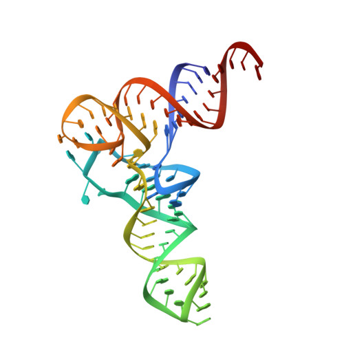
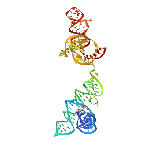
    T-box riboregulators are a class of cis-regulatory RNAs that govern the bacterial response to amino acid starvation by binding, decoding and reading the aminoacylation status of specific transfer RNAs. Here we provide a high-resolution crystal structure of a full-length T-box from Mycobacterium tuberculosis that explains tRNA decoding and aminoacylation sensing by this riboregulator. Overall, the T-box consists of decoding and aminoacylation sensing modules bridged by a rigid pseudoknot structure formed by the mid-region domains. Stem-I and the Stem-II S-turn assemble a claw-like decoding module, while the antiterminator, Stem-III, and the adjacent linker form a tightly interwoven aminoacylation sensing module. The uncharged tRNA is selectively recognized by an unexpected set of favorable contacts from the linker region in the aminoacylation sensing module. A complex structure with a charged tRNA mimic shows that the extra moiety dislodges the linker, which is indicative of the possible chain of events that lead to alternative base-pairing and altered expression output.


  • Organizational Affiliation
    • Department of Molecular Biology and Genetics, Ithaca, NY, USA.

Macromolecule Content 

  • Total Structure Weight: 79.38 kDa 
  • Atom Count: 5,154 
  • Modeled Residue Count: 239 
  • Deposited Residue Count: 243 
  • Unique nucleic acid chains: 2

Macromolecules

Find similar nucleic acids by:  (by identity cutoff) 
Entity ID: 1
MoleculeChains LengthOrganismImage
RNA (77-MER)A [auth B]77Mycobacterium tuberculosis
Sequence Annotations
Expand
Reference Sequence
Find similar nucleic acids by:  (by identity cutoff) 
Entity ID: 2
MoleculeChains LengthOrganismImage
RNA (166-MER)B [auth A]166Mycobacterium tuberculosis
Sequence Annotations
Expand
Reference Sequence

Small Molecules

Ligands 2 Unique
IDChains Name / Formula / InChI Key2D Diagram3D Interactions
K

Query on K


Download:Ideal Coordinates CCD File
E [auth B],
F [auth B],
P [auth A],
Q [auth A]
POTASSIUM ION
K
NPYPAHLBTDXSSS-UHFFFAOYSA-N
MG

Query on MG


Download:Ideal Coordinates CCD File
C [auth B]
D [auth B]
G [auth A]
H [auth A]
I [auth A]
C [auth B],
D [auth B],
G [auth A],
H [auth A],
I [auth A],
J [auth A],
K [auth A],
L [auth A],
M [auth A],
N [auth A],
O [auth A]
MAGNESIUM ION
Mg
JLVVSXFLKOJNIY-UHFFFAOYSA-N

Experimental Data & Validation

Experimental Data

  • Method: X-RAY DIFFRACTION
  • Resolution: 2.93 Å
  • R-Value Free:  0.235 (Depositor), 0.252 (DCC) 
  • R-Value Work:  0.198 (Depositor), 0.214 (DCC) 
  • R-Value Observed: 0.201 (Depositor) 
Space Group: P 41
Unit Cell:
Length ( Å )Angle ( ˚ )
a = 77.209α = 90
b = 77.209β = 90
c = 172.754γ = 90
Software Package:
Software NamePurpose
PHENIXrefinement
Aimlessdata scaling
PDB_EXTRACTdata extraction
XDSdata reduction
PHASERphasing

Structure Validation

View Full Validation Report


Entry History 

& Funding Information

Deposition Data


Funding OrganizationLocationGrant Number
National Institutes of Health/National Institute of General Medical Sciences (NIH/NIGMS)United StatesGM118174
National Institutes of Health/National Institute of General Medical Sciences (NIH/NIGMS)United StatesGM116632

Revision History  (Full details and data files)

  • Version 1.0: 2019-11-20
    Type: Initial release
  • Version 1.1: 2019-12-04
    Changes: Database references
  • Version 1.2: 2019-12-18
    Changes: Database references
  • Version 1.3: 2020-01-01
    Changes: Author supporting evidence
  • Version 1.4: 2024-03-13
    Changes: Data collection, Database references, Derived calculations