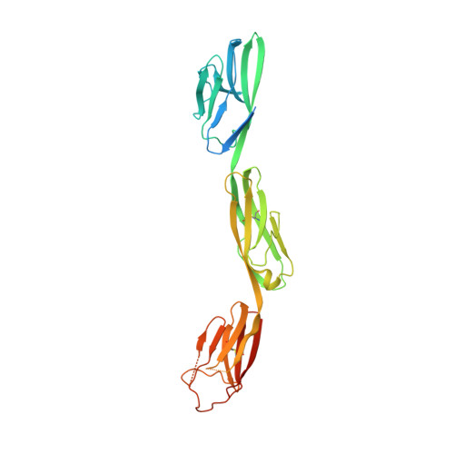
6U7N

Crystal structure of neurotrimin (NTM)


Experimental Data Snapshot

  • Method: X-RAY DIFFRACTION
  • Resolution: 3.32 Å
  • R-Value Free: 0.250 
  • R-Value Work: 0.229 
  • R-Value Observed: 0.230 

Starting Model: experimental
View more details

wwPDB Validation   3D Report Full Report


This is version 1.3 of the entry. See complete history


Literature

Highly Conserved Molecular Features in IgLONs Contrast Their Distinct Structural and Biological Outcomes.

Venkannagari, H.Kasper, J.M.Misra, A.Rush, S.A.Fan, S.Lee, H.Sun, H.Seshadrinathan, S.Machius, M.Hommel, J.D.Rudenko, G.

(2020) J Mol Biol 432: 5287-5303

  • DOI: https://doi.org/10.1016/j.jmb.2020.07.014
  • Primary Citation of Related Structures:  
    6U6T, 6U7N

  • PubMed Abstract: 

    Neuronal growth regulator 1 (NEGR1) and neurotrimin (NTM) are abundant cell-surface proteins found in the brain and form part of the IgLON (Immunoglobulin LSAMP, OBCAM, Neurotrimin) family. In humans, NEGR1 is implicated in obesity and mental disorders, while NTM is linked to intelligence and cognitive function. IgLONs dimerize homophilically and heterophilically, and they are thought to shape synaptic connections and neural circuits by acting in trans (spanning cellular junctions) and/or in cis (at the same side of a junction). Here, we reveal homodimeric structures of NEGR1 and NTM. They assemble into V-shaped complexes via their Ig1 domains, and disruption of the Ig1-Ig1 interface abolishes dimerization in solution. A hydrophobic ridge from one Ig1 domain inserts into a hydrophobic pocket from the opposing Ig1 domain producing an interaction interface that is highly conserved among IgLONs but remarkably plastic structurally. Given the high degree of sequence conservation at the interaction interface, we tested whether different IgLONs could elicit the same biological effect in vivo. In a small-scale study administering different soluble IgLONs directly into the brain and monitoring feeding, only NEGR1 altered food intake significantly. Taking NEGR1 as a prototype, our studies thus indicate that while IgLONs share a conserved mode of interaction and are able to bind each other as homomers and heteromers, they are structurally plastic and can exert unique biological action.


  • Organizational Affiliation

    Department of Pharmacology and Toxicology, University of Texas Medical Branch, Galveston, TX 77555, USA; Sealy Center for Structural Biology and Molecular Biophysics, University of Texas Medical Branch, Galveston, TX 77555, USA.


Macromolecules
Find similar proteins by:  (by identity cutoff)  |  3D Structure
Entity ID: 1
MoleculeChains Sequence LengthOrganismDetailsImage
Neurotrimin314Homo sapiensMutation(s): 0 
Gene Names: NTMIGLON2NTUNQ297/PRO337
UniProt & NIH Common Fund Data Resources
Find proteins for Q9P121 (Homo sapiens)
Explore Q9P121 
Go to UniProtKB:  Q9P121
PHAROS:  Q9P121
GTEx:  ENSG00000182667 
Entity Groups  
Sequence Clusters30% Identity50% Identity70% Identity90% Identity95% Identity100% Identity
UniProt GroupQ9P121
Glycosylation
Glycosylation Sites: 1Go to GlyGen: Q9P121-1
Sequence Annotations
Expand
  • Reference Sequence
Oligosaccharides

Help

Entity ID: 2
MoleculeChains Length2D Diagram Glycosylation3D Interactions
beta-D-mannopyranose-(1-4)-2-acetamido-2-deoxy-beta-D-glucopyranose-(1-4)-2-acetamido-2-deoxy-beta-D-glucopyranose
B
3N-Glycosylation
Glycosylation Resources
GlyTouCan:  G15407YE
GlyCosmos:  G15407YE
GlyGen:  G15407YE
Entity ID: 3
MoleculeChains Length2D Diagram Glycosylation3D Interactions
alpha-L-fucopyranose-(1-6)-2-acetamido-2-deoxy-beta-D-glucopyranose
C
2N/A
Glycosylation Resources
GlyTouCan:  G86851RC
GlyCosmos:  G86851RC
GlyGen:  G86851RC
Small Molecules
Ligands 2 Unique
IDChains Name / Formula / InChI Key2D Diagram3D Interactions
EDO
Query on EDO

Download Ideal Coordinates CCD File 
D [auth A],
E [auth A],
F [auth A]
1,2-ETHANEDIOL
C2 H6 O2
LYCAIKOWRPUZTN-UHFFFAOYSA-N
CL
Query on CL

Download Ideal Coordinates CCD File 
G [auth A]
H [auth A]
I [auth A]
J [auth A]
K [auth A]
G [auth A],
H [auth A],
I [auth A],
J [auth A],
K [auth A],
L [auth A],
M [auth A],
N [auth A]
CHLORIDE ION
Cl
VEXZGXHMUGYJMC-UHFFFAOYSA-M
Experimental Data & Validation

Experimental Data

  • Method: X-RAY DIFFRACTION
  • Resolution: 3.32 Å
  • R-Value Free: 0.250 
  • R-Value Work: 0.229 
  • R-Value Observed: 0.230 
  • Space Group: I 41
Unit Cell:
Length ( Å )Angle ( ˚ )
a = 106.049α = 90
b = 106.049β = 90
c = 227.819γ = 90
Software Package:
Software NamePurpose
PHENIXrefinement
HKL-2000data reduction
HKL-2000data scaling
PDB_EXTRACTdata extraction
PHENIXphasing

Structure Validation

View Full Validation Report


Entry History & Funding Information

Deposition Data


Funding OrganizationLocationGrant Number
National Institutes of Health/National Institute of Mental Health (NIH/NIMH)United StatesR01MH077303

Revision History  (Full details and data files)

  • Version 1.0: 2020-08-12
    Type: Initial release
  • Version 1.1: 2020-09-30
    Changes: Database references
  • Version 1.2: 2023-10-11
    Changes: Data collection, Database references, Refinement description
  • Version 1.3: 2024-10-23
    Changes: Structure summary