6U58 | pdb_00006u58

Toho1 Beta Lactamase Glu166Gln Mutant


Experimental Data Snapshot

  • Method: X-RAY DIFFRACTION
  • Resolution: 1.90 Å
  • R-Value Free: 
    0.180 (Depositor), 0.177 (DCC) 
  • R-Value Work: 
    0.150 (Depositor), 0.147 (DCC) 

  • Method: NEUTRON DIFFRACTION
  • Resolution: 1.89 Å
  • R-Value Free: 
    0.260 (Depositor) 
  • R-Value Work: 
    0.240 (Depositor) 

Starting Model: experimental
View more details

wwPDB Validation 3D Report Full Report

Validation slider image for 6U58

This is version 1.2 of the entry. See complete history

Literature

Probing the Role of the Conserved Residue Glu166 in a Class A Beta-Lactamase Using Neutron and X-ray Protein Crystallography

Langan, P.S.Sullivan, B.Weiss, K.L.Coates, L.

(2020) Acta Crystallogr D Biol Crystallogr 76: 118-123

  • DOI: https://doi.org/10.1107/S2059798319016334
  • Primary Citation Related Structures: 
    6U58

  • PubMed Abstract: 

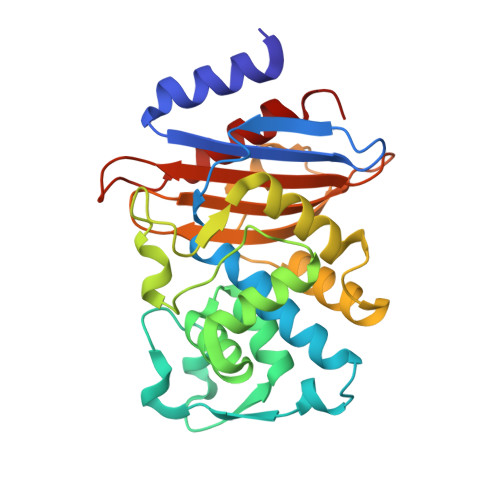
    The amino-acid sequence of the Toho-1 β-lactamase contains several conserved residues in the active site, including Ser70, Lys73, Ser130 and Glu166, some of which coordinate a catalytic water molecule. This catalytic water molecule is essential in the acylation and deacylation parts of the reaction mechanism through which Toho-1 inactivates specific antibiotics and provides resistance to its expressing bacterial strains. To investigate the function of Glu166 in the acylation part of the catalytic mechanism, neutron and X-ray crystallographic studies were performed on a Glu166Gln mutant. The structure of this class A β-lactamase mutant provides several insights into its previously reported reduced drug-binding kinetic rates. A joint refinement of both X-ray and neutron diffraction data was used to study the effects of the Glu166Gln mutation on the active site of Toho-1. This structure reveals that while the Glu166Gln mutation has a somewhat limited impact on the positions of the conserved amino acids within the active site, it displaces the catalytic water molecule from the active site. These subtle changes offer a structural explanation for the previously observed decreases in the binding of non-β-lactam inhibitors such as the recently developed diazobicyclooctane inhibitor avibactam.


  • Organizational Affiliation
    • Neutron Scattering Division, Oak Ridge National Laboratory, Oak Ridge, TN 37831, USA.

Macromolecule Content 

  • Total Structure Weight: 28.3 kDa 
  • Atom Count: 2,164 
  • Modeled Residue Count: 261 
  • Deposited Residue Count: 261 
  • Unique protein chains: 1

Macromolecules

Find similar proteins by:|  3D Structure
Entity ID: 1
MoleculeChains  Sequence LengthOrganismDetailsImage
Beta-lactamase261Escherichia coliMutation(s): 1 
Gene Names: bla CTX-M-56
EC: 3.5.2.6
UniProt
Find proteins for Q47066 (Escherichia coli)
Explore Q47066 
Go to UniProtKB:  Q47066
Entity Groups
Sequence Clusters30% Identity50% Identity70% Identity90% Identity95% Identity100% Identity
UniProt GroupQ47066
Sequence Annotations
Expand
Reference Sequence

Small Molecules

Experimental Data & Validation

Experimental Data

  • Method: X-RAY DIFFRACTION
  • Resolution: 1.90 Å
  • R-Value Free:  0.180 (Depositor), 0.177 (DCC) 
  • R-Value Work:  0.150 (Depositor), 0.147 (DCC) 
  • Method: NEUTRON DIFFRACTION
  • Resolution: 1.89 Å
  • R-Value Free:  0.260 (Depositor) 
  • R-Value Work:  0.240 (Depositor) 
Space Group: P 32 2 1
Unit Cell:
Length ( Å )Angle ( ˚ )
a = 73.543α = 90
b = 73.543β = 90
c = 99.721γ = 120
Software Package:
Software NamePurpose
PHENIXrefinement
CrysalisProdata reduction
Mantiddata reduction

Structure Validation

View Full Validation Report


Entry History 

Deposition Data

Revision History  (Full details and data files)

  • Version 1.0: 2020-02-19
    Type: Initial release
  • Version 1.1: 2024-03-13
    Changes: Data collection, Database references
  • Version 1.2: 2024-04-03
    Changes: Refinement description