The 3-His Metal Coordination Site Promotes the Coupling of Oxygen Activation to Cysteine Oxidation in Cysteine Dioxygenase.
Forbes, D.L., Meneely, K.M., Chilton, A.S., Lamb, A.L., Ellis, H.R.(2020) Biochemistry 59: 2022-2031
- PubMed: 32368901 Search on PubMedSearch on PubMed Central
- DOI: https://doi.org/10.1021/acs.biochem.9b01085
- Primary Citation Related Structures:
6U4L, 6U4S, 6U4V - PubMed Abstract:
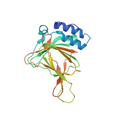
Cysteine dioxygenase (CDO) structurally resembles cupin enzymes that use a 3-His/1-Glu coordination scheme. However, the glutamate ligand is substituted with a cysteine (Cys93) residue, which forms a thioether bond with tyrosine (Tyr157) under physiological conditions. The reversion variant, C93E CDO, was generated in order to reestablish the more common 3-His/1-Glu metal ligands of the cupin superfamily. This variant provides a framework for testing the structural and functional significance of Cys93 and the cross-link in CDO. Although dioxygen consumption was observed with C93E CDO, it was not coupled with l-cysteine oxidation. Substrate analogues (d-cysteine, cysteamine, and 3-mercaptopropionate) were not viable substrates for the C93E CDO variant, although they showed variable coordinations to the iron center. The structures of C93E and cross-linked and non-cross-linked wild-type CDO were solved by X-ray crystallography to 1.91, 2.49, and 2.30 Å, respectively. The C93E CDO variant had similar overall structural properties compared to cross-linked CDO; however, the iron was coordinated by a 3-His/1-Glu geometry, leaving only two coordination sites available for dioxygen and bidentate l-cysteine binding. The hydroxyl group of Tyr157 shifted in both non-cross-linked and C93E CDO, and this displacement prevented the residue from participating in substrate stabilization. Based on these results, the divergence of the metal center of cysteine dioxygenase from the 3-His/1-Glu geometry seen with many cupin enzymes was essential for effective substrate binding. The substitution of Glu with Cys in CDO allows for a third coordination site on the iron for bidentate cysteine and monodentate oxygen binding.
- The Department of Chemistry and Biochemistry, Auburn University, Auburn, Alabama 36849, United States.
Organizational Affiliation: