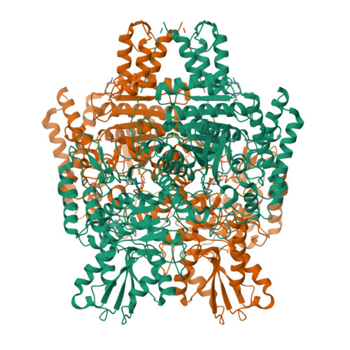
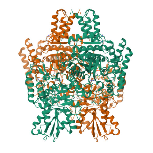
6U3J

Structure of the 2-oxoadipate dehydrogenase DHTKD1


Experimental Data Snapshot

  • Method: X-RAY DIFFRACTION
  • Resolution: 2.25 Å
  • R-Value Free: 0.241 
  • R-Value Work: 0.209 
  • R-Value Observed: 0.211 

Starting Model: experimental
View more details

wwPDB Validation   3D Report Full Report


Ligand Structure Quality Assessment 


This is version 1.3 of the entry. See complete history


Literature

Inhibition and Crystal Structure of the Human DHTKD1-Thiamin Diphosphate Complex.

Leandro, J.Khamrui, S.Wang, H.Suebsuwong, C.Nemeria, N.S.Huynh, K.Moustakim, M.Secor, C.Wang, M.Dodatko, T.Stauffer, B.Wilson, C.G.Yu, C.Arkin, M.R.Jordan, F.Sanchez, R.DeVita, R.J.Lazarus, M.B.Houten, S.M.

(2020) ACS Chem Biol 15: 2041-2047

  • DOI: https://doi.org/10.1021/acschembio.0c00114
  • Primary Citation of Related Structures:  
    6U3J

  • PubMed Abstract: 

    DHTKD1 is the E1 component of the 2-oxoadipate dehydrogenase complex, which is an enzyme involved in the catabolism of (hydroxy-)lysine and tryptophan. Mutations in DHTKD1 have been associated with 2-aminoadipic and 2-oxoadipic aciduria, Charcot-Marie-Tooth disease type 2Q and eosinophilic esophagitis, but the pathophysiology of these clinically distinct disorders remains elusive. Here, we report the identification of adipoylphosphonic acid and tenatoprazole as DHTKD1 inhibitors using targeted and high throughput screening, respectively. We furthermore elucidate the DHTKD1 crystal structure with thiamin diphosphate bound at 2.25 Å. We also report the impact of 10 disease-associated missense mutations on DHTKD1. Whereas the majority of the DHTKD1 variants displayed impaired folding or reduced thermal stability in combination with absent or reduced enzyme activity, three variants showed no abnormalities. Our work provides chemical and structural tools for further understanding of the function of DHTKD1 and its role in several human pathologies.


  • Organizational Affiliation

    Department of Genetics and Genomic Sciences, Icahn School of Medicine at Mount Sinai, New York, New York 10029, United States.


Macromolecules
Find similar proteins by:  (by identity cutoff)  |  3D Structure
Entity ID: 1
MoleculeChains Sequence LengthOrganismDetailsImage
2-oxoglutarate dehydrogenase E1 component DHKTD1, mitochondrial
A, B
904Homo sapiensMutation(s): 0 
Gene Names: DHTKD1KIAA1630
EC: 1.2.4.2 (PDB Primary Data), 1.2.4 (UniProt)
UniProt & NIH Common Fund Data Resources
Find proteins for Q96HY7 (Homo sapiens)
Explore Q96HY7 
Go to UniProtKB:  Q96HY7
PHAROS:  Q96HY7
GTEx:  ENSG00000181192 
Entity Groups  
Sequence Clusters30% Identity50% Identity70% Identity90% Identity95% Identity100% Identity
UniProt GroupQ96HY7
Sequence Annotations
Expand
  • Reference Sequence
Experimental Data & Validation

Experimental Data

  • Method: X-RAY DIFFRACTION
  • Resolution: 2.25 Å
  • R-Value Free: 0.241 
  • R-Value Work: 0.209 
  • R-Value Observed: 0.211 
  • Space Group: C 1 2 1
Unit Cell:
Length ( Å )Angle ( ˚ )
a = 332.314α = 90
b = 72.612β = 91.87
c = 79.675γ = 90
Software Package:
Software NamePurpose
PHENIXrefinement
MOSFLMdata reduction
Aimlessdata scaling
PHASERphasing
PDB_EXTRACTdata extraction

Structure Validation

View Full Validation Report


Ligand Structure Quality Assessment 


Entry History & Funding Information

Deposition Data


Funding OrganizationLocationGrant Number
National Institutes of Health/National Institute of General Medical Sciences (NIH/NIGMS)United StatesGM124838

Revision History  (Full details and data files)

  • Version 1.0: 2020-07-22
    Type: Initial release
  • Version 1.1: 2020-07-29
    Changes: Database references, Derived calculations
  • Version 1.2: 2020-09-02
    Changes: Database references
  • Version 1.3: 2023-10-11
    Changes: Data collection, Database references, Refinement description