6U2T

Crystal structure of the T-state of maize C4-phosphoenolpyruvate carboxylase in complex with malate


Experimental Data Snapshot

  • Method: X-RAY DIFFRACTION
  • Resolution: 2.80 Å
  • R-Value Free: 0.226 
  • R-Value Work: 0.183 
  • R-Value Observed: 0.185 

Starting Model: experimental
View more details

wwPDB Validation   3D Report Full Report


This is version 1.2 of the entry. See complete history


Literature

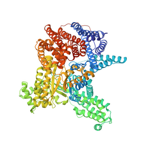
Two new T-state crystal structures of maize C 4 -phosphoenolpyruvate carboxylase reveal and suggest novel structural features of the allosteric regulation and carboxylation step.

Carrizosa-Carbajal, E.I.Gonzalez-Segura, L.Munoz-Clares, R.A.

(2024) Int J Biol Macromol : 135134-135134

  • DOI: https://doi.org/10.1016/j.ijbiomac.2024.135134
  • Primary Citation of Related Structures:  
    6U2T, 6V3O

  • PubMed Abstract: 

    To get a deeper understanding of the structural bases of the allosteric transition between T and R states of plant and bacterial phosphoenolpyruvate carboxylases (PEPCs), we obtained the first T-state crystal structures of the maize photosynthetic PEPC (ZmPEPC-C 4 ) and exhaustively compared them with the previously reported R-state ZmPEPC-C 4 and other T-state structures. We identified previously unrecognized significant conformational changes in the T state: that of the α8-α9 loop, which connects the two kinds of activator allosteric sites with the active site, the conversion of the α30 helix into a 3 10 helix, leading to the disorganization of the active site lid and activators allosteric sites, and the closure of the inhibitor allosteric-site lid. Additionally, we identified previously overlooked, highly conserved residues of potential interest in the allosteric transition, including two histidines whose protonation might stabilize the T state. The crystal structures reported here also suggest similar tetrameric quaternary arrangements of PEPC enzymes in the R and T states, and the location of the bicarbonate binding site, as well as the conformational changes required for the carboxylation step. Our findings and working hypothesis advance the understanding of the structural features of the allosteric PEPC enzymes and provide a foundation for future experiments.


  • Organizational Affiliation

    Departamento de Bioquímica, Facultad de Química, Universidad Nacional Autónoma de México, Ciudad Universitaria, 04510 Ciudad de México, Mexico.


Macromolecules
Find similar proteins by:  (by identity cutoff)  |  3D Structure
Entity ID: 1
MoleculeChains Sequence LengthOrganismDetailsImage
Phosphoenolpyruvate carboxylase
A, B, C, D
970Zea maysMutation(s): 0 
Gene Names: ppc1
EC: 4.1.1.31
UniProt
Find proteins for P04711 (Zea mays)
Explore P04711 
Go to UniProtKB:  P04711
Entity Groups  
Sequence Clusters30% Identity50% Identity70% Identity90% Identity95% Identity100% Identity
UniProt GroupP04711
Sequence Annotations
Expand
  • Reference Sequence
Small Molecules
Ligands 3 Unique
IDChains Name / Formula / InChI Key2D Diagram3D Interactions
MLT
Query on MLT

Download Ideal Coordinates CCD File 
E [auth A],
H [auth B],
L [auth C],
P [auth D]
D-MALATE
C4 H6 O5
BJEPYKJPYRNKOW-UWTATZPHSA-N
SO4
Query on SO4

Download Ideal Coordinates CCD File 
F [auth A]
G [auth A]
I [auth B]
J [auth B]
K [auth B]
F [auth A],
G [auth A],
I [auth B],
J [auth B],
K [auth B],
M [auth C],
N [auth C],
O [auth C],
Q [auth D],
R [auth D],
S [auth D]
SULFATE ION
O4 S
QAOWNCQODCNURD-UHFFFAOYSA-L
GOL
Query on GOL

Download Ideal Coordinates CCD File 
T [auth D]GLYCEROL
C3 H8 O3
PEDCQBHIVMGVHV-UHFFFAOYSA-N
Experimental Data & Validation

Experimental Data

  • Method: X-RAY DIFFRACTION
  • Resolution: 2.80 Å
  • R-Value Free: 0.226 
  • R-Value Work: 0.183 
  • R-Value Observed: 0.185 
  • Space Group: P 21 21 21
Unit Cell:
Length ( Å )Angle ( ˚ )
a = 103.94α = 90
b = 140.931β = 90
c = 376.401γ = 90
Software Package:
Software NamePurpose
PHENIXrefinement
SCALAdata scaling
PHASERphasing
PDB_EXTRACTdata extraction
XDSdata reduction

Structure Validation

View Full Validation Report


Entry History 

Deposition Data

Revision History  (Full details and data files)

  • Version 1.0: 2020-09-09
    Type: Initial release
  • Version 1.1: 2023-10-11
    Changes: Data collection, Database references, Refinement description
  • Version 1.2: 2024-09-11
    Changes: Database references