6TYX

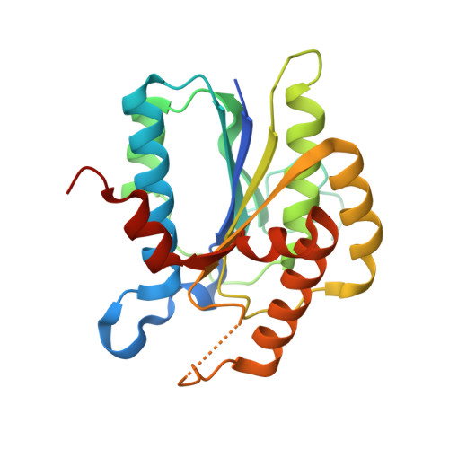
Structure of Ku80 von Willebrand domain S229A mutant complexed with XLF Ku Binding Motif


Experimental Data Snapshot

  • Method: X-RAY DIFFRACTION
  • Resolution: 1.90 Å
  • R-Value Free: 0.225 
  • R-Value Work: 0.185 
  • R-Value Observed: 0.187 

Starting Model: experimental
View more details

wwPDB Validation   3D Report Full Report

Currently 6TYX does not have a validation slider image.


This is version 1.1 of the entry. See complete history


Literature

Ligand binding characteristics of the Ku80 von Willebrand domain.

Kim, K.Min, J.Kirby, T.W.Gabel, S.A.Pedersen, L.C.London, R.E.

(2019) DNA Repair (Amst) 85: 102739-102739

  • DOI: https://doi.org/10.1016/j.dnarep.2019.102739
  • Primary Citation of Related Structures:  
    6TYT, 6TYU, 6TYV, 6TYW, 6TYX, 6TYZ

  • PubMed Abstract: 

    The N-terminal von Willebrand domain of Ku80 supports interactions with a Ku binding motif (KBM) that has been identified in at least three other DNA repair proteins: the non-homologous end joining (NHEJ) scaffold APLF, the modulator of retrovirus infection, MRI, and the Werner syndrome protein (WRN). A second, more recently identified Ku binding motif present in XLF and several other proteins (KBMX) has also been reported to interact with this domain. The isolated Ku80 von Willebrand antigen domain (vWA) from Xenopus laevis has a sequence that is 60% identical with the human domain, is readily expressed and has been used to investigate these interactions. Structural characterization of the complexes formed with the KBM motifs in human APLF, MRI, and WRN identify a conserved binding site that is consistent with previously-reported mutational studies. In contrast with the KBM binding site, structural studies indicate that the KBMX site is occluded by a distorted helix. Fluorescence polarization and 19 F NMR studies of a fluorinated XLF C-terminal peptide failed to indicate any interaction with the frog vWA. It was hypothesized that availability of this binding site is conditional, i.e., dependent on specific experimental conditions or other repair factors to make the site available for binding. Modulating the fraction of KBMX-accessible binding site mutationally demonstrated that the more open site is capable of binding the KBMX XLF motif peptide. It is suggested that the conditional nature of KBMX binding limits formation of non-productive complexes so that activation-dependent site availability can more optimally support advancing the synapsis process.


  • Organizational Affiliation

    Genome Integrity and Structural Biology Laboratory, National Institute of Environment and Health Sciences, National Institutes of Health, Research Triangle Park, NC, 27709, USA.


Macromolecules
Find similar proteins by:  (by identity cutoff)  |  3D Structure
Entity ID: 1
MoleculeChains Sequence LengthOrganismDetailsImage
X-ray repair cross-complementing protein 5
A, B
231Xenopus laevisMutation(s): 2 
Gene Names: XELAEV_18044412mg
EC: 3.6.4
UniProt
Find proteins for Q6DDS9 (Xenopus laevis)
Explore Q6DDS9 
Go to UniProtKB:  Q6DDS9
Entity Groups  
Sequence Clusters30% Identity50% Identity70% Identity90% Identity95% Identity100% Identity
UniProt GroupQ6DDS9
Sequence Annotations
Expand
  • Reference Sequence

Find similar proteins by:  Sequence   |   3D Structure  

Entity ID: 2
MoleculeChains Sequence LengthOrganismDetailsImage
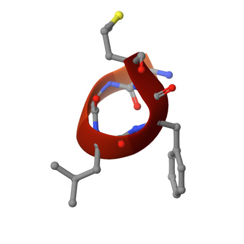
LYS-GLY-LEU-PHE-MET
C, D
18Xenopus laevisMutation(s): 0 
UniProt
Find proteins for A0A1L8ENT6 (Xenopus laevis)
Explore A0A1L8ENT6 
Go to UniProtKB:  A0A1L8ENT6
Entity Groups  
Sequence Clusters30% Identity50% Identity70% Identity90% Identity95% Identity100% Identity
UniProt GroupA0A1L8ENT6
Sequence Annotations
Expand
  • Reference Sequence
Experimental Data & Validation

Experimental Data

  • Method: X-RAY DIFFRACTION
  • Resolution: 1.90 Å
  • R-Value Free: 0.225 
  • R-Value Work: 0.185 
  • R-Value Observed: 0.187 
  • Space Group: P 1 21 1
Unit Cell:
Length ( Å )Angle ( ˚ )
a = 44.798α = 90
b = 71.605β = 98.309
c = 74.6γ = 90
Software Package:
Software NamePurpose
PHENIXrefinement
HKL-2000data reduction
HKL-2000data scaling
PHASERphasing

Structure Validation

View Full Validation Report

Currently 6TYX does not have a validation slider image.


Entry History & Funding Information

Deposition Data


Funding OrganizationLocationGrant Number
National Institutes of Health/National Institute of Environmental Health Sciences (NIH/NIEHS)United StatesZIA-ES102645

Revision History  (Full details and data files)

  • Version 1.0: 2019-11-27
    Type: Initial release
  • Version 1.1: 2023-10-11
    Changes: Data collection, Database references, Refinement description