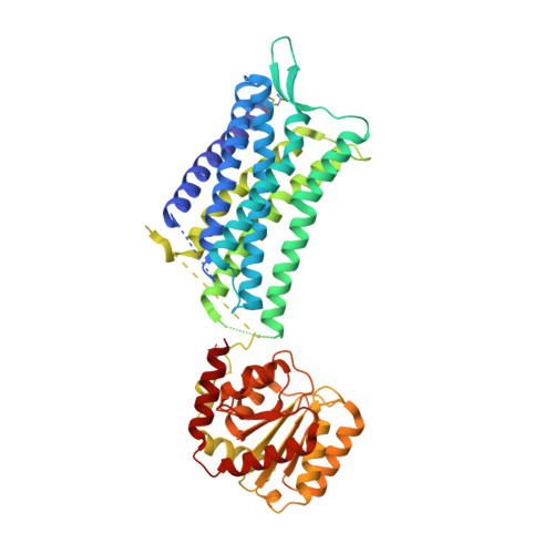
6TPK

Crystal structure of the human oxytocin receptor


Experimental Data Snapshot

  • Method: X-RAY DIFFRACTION
  • Resolution: 3.20 Å
  • R-Value Free: 0.266 
  • R-Value Work: 0.252 
  • R-Value Observed: 0.252 

wwPDB Validation   3D Report Full Report


Ligand Structure Quality Assessment 


This is version 1.2 of the entry. See complete history


Literature

Crystal structure of the human oxytocin receptor.

Waltenspuhl, Y.Schoppe, J.Ehrenmann, J.Kummer, L.Pluckthun, A.

(2020) Sci Adv 6: eabb5419-eabb5419

  • DOI: https://doi.org/10.1126/sciadv.abb5419
  • Primary Citation of Related Structures:  
    6TPK

  • PubMed Abstract: 

    The peptide hormone oxytocin modulates socioemotional behavior and sexual reproduction via the centrally expressed oxytocin receptor (OTR) across several species. Here, we report the crystal structure of human OTR in complex with retosiban, a nonpeptidic antagonist developed as an oral drug for the prevention of preterm labor. Our structure reveals insights into the detailed interactions between the G protein-coupled receptor (GPCR) and an OTR-selective antagonist. The observation of an extrahelical cholesterol molecule, binding in an unexpected location between helices IV and V, provides a structural rationale for its allosteric effect and critical influence on OTR function. Furthermore, our structure in combination with experimental data allows the identification of a conserved neurohypophyseal receptor-specific coordination site for Mg 2+ that acts as potent, positive allosteric modulator for agonist binding. Together, these results further our molecular understanding of the oxytocin/vasopressin receptor family and will facilitate structure-guided development of new therapeutics.


  • Organizational Affiliation

    Department of Biochemistry, University of Zürich, Winterthurerstrasse 190, CH-8057 Zürich, Switzerland.


Macromolecules
Find similar proteins by:  (by identity cutoff)  |  3D Structure
Entity ID: 1
MoleculeChains Sequence LengthOrganismDetailsImage
Oxytocin receptor502Homo sapiensMutation(s): 0 
Gene Names: OXTR
Membrane Entity: Yes 
UniProt & NIH Common Fund Data Resources
Find proteins for P30559 (Homo sapiens)
Explore P30559 
Go to UniProtKB:  P30559
PHAROS:  P30559
GTEx:  ENSG00000180914 
Entity Groups  
Sequence Clusters30% Identity50% Identity70% Identity90% Identity95% Identity100% Identity
UniProt GroupP30559
Sequence Annotations
Expand
  • Reference Sequence
Small Molecules
Ligands 3 Unique
IDChains Name / Formula / InChI Key2D Diagram3D Interactions
NU2 (Subject of Investigation/LOI)
Query on NU2

Download Ideal Coordinates CCD File 
B [auth A](3~{R},6~{R})-6-[(2~{S})-butan-2-yl]-3-(2,3-dihydro-1~{H}-inden-2-yl)-1-[(1~{R})-1-(2-methyl-1,3-oxazol-4-yl)-2-morpholin-4-yl-2-oxidanylidene-ethyl]piperazine-2,5-dione
C27 H34 N4 O5
PLVGDGRBPMVYPB-FDUHJNRSSA-N
CLR (Subject of Investigation/LOI)
Query on CLR

Download Ideal Coordinates CCD File 
C [auth A]CHOLESTEROL
C27 H46 O
HVYWMOMLDIMFJA-DPAQBDIFSA-N
PEG
Query on PEG

Download Ideal Coordinates CCD File 
D [auth A]DI(HYDROXYETHYL)ETHER
C4 H10 O3
MTHSVFCYNBDYFN-UHFFFAOYSA-N
Modified Residues  1 Unique
IDChains TypeFormula2D DiagramParent
YCM
Query on YCM
A
L-PEPTIDE LINKINGC5 H10 N2 O3 SCYS
Binding Affinity Annotations 
IDSourceBinding Affinity
NU2 BindingDB:  6TPK Ki: min: 1, max: 4.1 (nM) from 2 assay(s)
Experimental Data & Validation

Experimental Data

  • Method: X-RAY DIFFRACTION
  • Resolution: 3.20 Å
  • R-Value Free: 0.266 
  • R-Value Work: 0.252 
  • R-Value Observed: 0.252 
  • Space Group: C 2 2 21
Unit Cell:
Length ( Å )Angle ( ˚ )
a = 76.987α = 90
b = 78.858β = 90
c = 283.486γ = 90
Software Package:
Software NamePurpose
Aimlessdata scaling
PHENIXrefinement
PDB_EXTRACTdata extraction
XDSdata reduction
PHASERphasing

Structure Validation

View Full Validation Report


Ligand Structure Quality Assessment 


Entry History & Funding Information

Deposition Data


Funding OrganizationLocationGrant Number
Swiss National Science FoundationSwitzerland31003A_153143
Swiss National Science FoundationSwitzerland31003A_182334

Revision History  (Full details and data files)

  • Version 1.0: 2020-08-05
    Type: Initial release
  • Version 1.1: 2020-08-19
    Changes: Structure summary
  • Version 1.2: 2020-09-02
    Changes: Database references