6TGK | pdb_00006tgk

Domain swapped E6AP C-lobe dimer


Experimental Data Snapshot

  • Method: X-RAY DIFFRACTION
  • Resolution: 1.30 Å
  • R-Value Free: 
    0.207 (Depositor), 0.216 (DCC) 
  • R-Value Work: 
    0.178 (Depositor), 0.188 (DCC) 
  • R-Value Observed: 
    0.179 (Depositor) 

Starting Model: experimental
View more details

wwPDB Validation 3D Report Full Report

Validation slider image for 6TGK

This is version 1.2 of the entry. See complete history

Literature

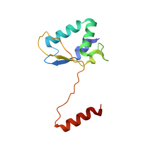
Crystal structure of the catalytic C-lobe of the HECT-type ubiquitin ligase E6AP.

Ries, L.K.Liess, A.K.L.Feiler, C.G.Spratt, D.E.Lowe, E.D.Lorenz, S.

(2020) Protein Sci 29: 1550-1554

  • DOI: https://doi.org/10.1002/pro.3832
  • Primary Citation Related Structures: 
    6TGK

  • PubMed Abstract: 

    The HECT-type ubiquitin ligase E6AP (UBE3A) is critically involved in several neurodevelopmental disorders and human papilloma virus-induced cervical tumorigenesis; the structural mechanisms underlying the activity of this crucial ligase, however, are incompletely understood. Here, we report a crystal structure of the C-terminal lobe ("C-lobe") of the catalytic domain of E6AP that reveals two molecules in a domain-swapped, dimeric arrangement. Interestingly, the molecular hinge that enables this structural reorganization with respect to the monomeric fold coincides with the active-site region. While such dimerization is unlikely to occur in the context of full-length E6AP, we noticed a similar domain swap in a crystal structure of the isolated C-lobe of another HECT-type ubiquitin ligase, HERC6. This may point to conformational strain in the active-site region of HECT-type ligases with possible implications for catalysis. SIGNIFICANCE STATEMENT: The HECT-type ubiquitin ligase E6AP has key roles in human papilloma virus-induced cervical tumorigenesis and certain neurodevelopmental disorders. Here, we present a crystal structure of the C-terminal, catalytic lobe of E6AP, providing basic insight into the conformational properties of this functionally critical region of HECT-type ligases.


  • Organizational Affiliation
    • Rudolf Virchow Center for Experimental Biomedicine, University of Würzburg, Würzburg, Germany.

Macromolecule Content 

  • Total Structure Weight: 12.22 kDa 
  • Atom Count: 1,051 
  • Modeled Residue Count: 105 
  • Deposited Residue Count: 105 
  • Unique protein chains: 1

Macromolecules

Find similar proteins by:|  3D Structure
Entity ID: 1
MoleculeChains  Sequence LengthOrganismDetailsImage
Ubiquitin-protein ligase E3AA [auth C]105Homo sapiensMutation(s): 0 
Gene Names: UBE3AE6APEPVE6APHPVE6A
EC: 2.3.2.26
UniProt & NIH Common Fund Data Resources
Find proteins for Q05086 (Homo sapiens)
Explore Q05086 
Go to UniProtKB:  Q05086
PHAROS:  Q05086
GTEx:  ENSG00000114062 
Entity Groups
Sequence Clusters30% Identity50% Identity70% Identity90% Identity95% Identity100% Identity
UniProt GroupQ05086
Sequence Annotations
Expand
Reference Sequence

Experimental Data & Validation

Experimental Data

  • Method: X-RAY DIFFRACTION
  • Resolution: 1.30 Å
  • R-Value Free:  0.207 (Depositor), 0.216 (DCC) 
  • R-Value Work:  0.178 (Depositor), 0.188 (DCC) 
  • R-Value Observed: 0.179 (Depositor) 
Space Group: I 4 2 2
Unit Cell:
Length ( Å )Angle ( ˚ )
a = 72.28α = 90
b = 72.28β = 90
c = 98.974γ = 90
Software Package:
Software NamePurpose
REFMACrefinement
Aimlessdata scaling
PDB_EXTRACTdata extraction
Aimlessdata reduction
PHASERphasing

Structure Validation

View Full Validation Report


Entry History 

& Funding Information

Deposition Data


Funding OrganizationLocationGrant Number
German Research FoundationGermanyLO 2003/1-1

Revision History  (Full details and data files)

  • Version 1.0: 2020-02-26
    Type: Initial release
  • Version 1.1: 2020-06-10
    Changes: Database references
  • Version 1.2: 2024-01-24
    Changes: Data collection, Database references, Derived calculations, Refinement description