6TCJ | pdb_00006tcj

Crystal structure of the BCL6 BTB domain in complex with a hybrid BTB-binding (HBP) peptide


Experimental Data Snapshot

  • Method: X-RAY DIFFRACTION
  • Resolution: 2.13 Å
  • R-Value Free: 
    0.249 (Depositor), 0.249 (DCC) 
  • R-Value Work: 
    0.199 (Depositor), 0.206 (DCC) 
  • R-Value Observed: 
    0.202 (Depositor) 

Starting Model: experimental
View more details

wwPDB Validation 3D Report Full Report

Validation slider image for 6TCJ

This is version 1.2 of the entry. See complete history

Literature

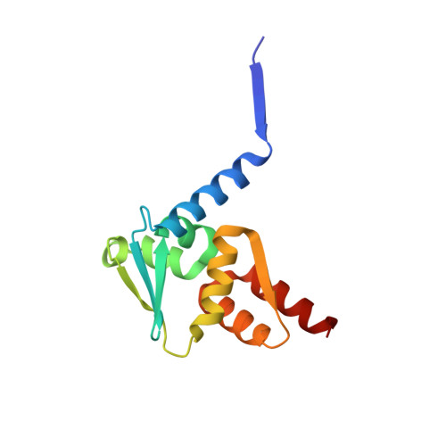
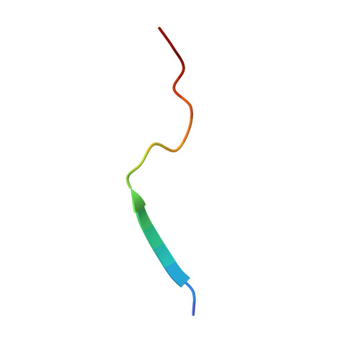
Structural basis of Apt48 inhibition of the BCL6 BTB domain.

Zacharchenko, T.Kalverda, A.P.Wright, S.C.

(2021) Structure 

  • DOI: https://doi.org/10.1016/j.str.2021.10.010
  • Primary Citation Related Structures: 
    6TBT, 6TCJ

  • PubMed Abstract: 

    B cell lymphoma 6 (BCL6) is a transcriptional repressor that is deregulated in diffuse large B cell lymphoma, and the peptide aptamer, Apt48, inhibits BCL6 by an unknown mechanism. We report the crystal structure of BCL6 in complex with an Apt48 peptide, and show that Apt48 binds to a therapeutically uncharacterized region at the bottom of the BCL6 BTB domain. We show that the corepressor binding site of the BTB domain may be divided conceptually into two low-affinity, peptide-binding regions. An upper region, the lateral groove, binds peptides in robust three-dimensional conformations, whereas a lower binding site is permissive to less-specific interactions. We show that, even with little sequence specificity, the interactions of the lower region are required for the high-affinity binding of the SMRT corepressor and other peptides to the BTB domain. This has relevance for the design of new BCL6 inhibitors and for understanding the evolution of corepressor interactions with the BTB domain.


  • Organizational Affiliation
    • School of Biology and the Astbury Centre for Structural Molecular Biology, University of Leeds, Leeds LS2 9JT, UK.

Macromolecule Content 

  • Total Structure Weight: 32.91 kDa 
  • Atom Count: 2,354 
  • Modeled Residue Count: 280 
  • Deposited Residue Count: 286 
  • Unique protein chains: 2

Macromolecules

Find similar proteins by:|  3D Structure
Entity ID: 1
MoleculeChains  Sequence LengthOrganismDetailsImage
B-cell lymphoma 6 protein
A, B
126Homo sapiensMutation(s): 3 
Gene Names: BCL6BCL5LAZ3ZBTB27ZNF51
UniProt & NIH Common Fund Data Resources
Find proteins for P41182 (Homo sapiens)
Explore P41182 
Go to UniProtKB:  P41182
PHAROS:  P41182
GTEx:  ENSG00000113916 
Entity Groups
Sequence Clusters30% Identity50% Identity70% Identity90% Identity95% Identity100% Identity
UniProt GroupP41182
Sequence Annotations
Expand
Reference Sequence
Find similar proteins by:  Sequence   |   3D Structure  
Entity ID: 2
MoleculeChains  Sequence LengthOrganismDetailsImage
Hybrid BTB-binding (HBP) peptide
C, D
17synthetic constructMutation(s): 0 

Small Molecules

Ligands 1 Unique
IDChains Name / Formula / InChI Key2D Diagram3D Interactions
NA

Query on NA


Download:Ideal Coordinates CCD File
E [auth B]SODIUM ION
Na
FKNQFGJONOIPTF-UHFFFAOYSA-N

Experimental Data & Validation

Experimental Data

  • Method: X-RAY DIFFRACTION
  • Resolution: 2.13 Å
  • R-Value Free:  0.249 (Depositor), 0.249 (DCC) 
  • R-Value Work:  0.199 (Depositor), 0.206 (DCC) 
  • R-Value Observed: 0.202 (Depositor) 
Space Group: P 1 21 1
Unit Cell:
Length ( Å )Angle ( ˚ )
a = 39.657α = 90
b = 47.3β = 94.34
c = 76.37γ = 90
Software Package:
Software NamePurpose
REFMACrefinement
XDSdata reduction
XSCALEdata scaling
PHASERphasing

Structure Validation

View Full Validation Report


Entry History 

& Funding Information

Deposition Data


Funding OrganizationLocationGrant Number
Kay Kendall Leukaemia FundUnited KingdomKKLF1047

Revision History  (Full details and data files)

  • Version 1.0: 2021-10-13
    Type: Initial release
  • Version 1.1: 2021-11-24
    Changes: Data collection, Database references
  • Version 1.2: 2024-01-24
    Changes: Data collection, Refinement description