6TCH | pdb_00006tch

Binary complex of 14-3-3 sigma and a high-affinity non-canonical 9-mer peptide binder


Experimental Data Snapshot

  • Method: X-RAY DIFFRACTION
  • Resolution: 1.80 Å
  • R-Value Free: 
    0.184 (Depositor), 0.184 (DCC) 
  • R-Value Work: 
    0.148 (Depositor), 0.149 (DCC) 
  • R-Value Observed: 
    0.150 (Depositor) 

Starting Model: experimental
View more details

wwPDB Validation 3D Report Full Report

Validation slider image for 6TCH

This is version 2.1 of the entry. See complete history

Literature

Ultra-large chemical libraries for the discovery of high-affinity peptide binders.

Quartararo, A.J.Gates, Z.P.Somsen, B.A.Hartrampf, N.Ye, X.Shimada, A.Kajihara, Y.Ottmann, C.Pentelute, B.L.

(2020) Nat Commun 11: 3183-3183

  • DOI: https://doi.org/10.1038/s41467-020-16920-3
  • Primary Citation Related Structures: 
    6TCH

  • PubMed Abstract: 

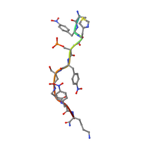
    High-diversity genetically-encoded combinatorial libraries (10 8 -10 13 members) are a rich source of peptide-based binding molecules, identified by affinity selection. Synthetic libraries can access broader chemical space, but typically examine only ~ 10 6 compounds by screening. Here we show that in-solution affinity selection can be interfaced with nano-liquid chromatography-tandem mass spectrometry peptide sequencing to identify binders from fully randomized synthetic libraries of 10 8 members-a 100-fold gain in diversity over standard practice. To validate this approach, we show that binders to a monoclonal antibody are identified in proportion to library diversity, as diversity is increased from 10 6 -10 8 . These results are then applied to the discovery of p53-like binders to MDM2, and to a family of 3-19 nM-affinity, α/β-peptide-based binders to 14-3-3. An X-ray structure of one of these binders in complex with 14-3-3σ is determined, illustrating the role of β-amino acids in facilitating a key binding contact.


  • Organizational Affiliation
    • Department of Chemistry, Massachusetts Institute of Technology, Cambridge, MA, 02139, USA.

Macromolecule Content 

  • Total Structure Weight: 28.12 kDa 
  • Atom Count: 2,375 
  • Modeled Residue Count: 246 
  • Deposited Residue Count: 248 
  • Unique protein chains: 2

Macromolecules

Find similar proteins by:|  3D Structure
Entity ID: 1
MoleculeChains  Sequence LengthOrganismDetailsImage
14-3-3 protein sigmaA [auth B]236Homo sapiensMutation(s): 0 
Gene Names: SFNHME1
UniProt & NIH Common Fund Data Resources
Find proteins for P31947 (Homo sapiens)
Explore P31947 
Go to UniProtKB:  P31947
PHAROS:  P31947
GTEx:  ENSG00000175793 
Entity Groups
Sequence Clusters30% Identity50% Identity70% Identity90% Identity95% Identity100% Identity
UniProt GroupP31947
Sequence Annotations
Expand
Reference Sequence
Find similar proteins by:  Sequence   |   3D Structure  
Entity ID: 2
MoleculeChains  Sequence LengthOrganismDetailsImage
DLY-NVA-PPN-KCJ-SEP-PPN-B3S-BAL-PPN-LYSB [auth A]12synthetic constructMutation(s): 0 

Small Molecules

Modified Residues  3 Unique
IDChains TypeFormula2D DiagramParent
B3S
Query on B3S
B [auth A]D-PEPTIDE LINKINGC4 H9 N O3

--

BAL
Query on BAL
B [auth A]PEPTIDE-LIKEC3 H7 N O2

--

DLY
Query on DLY
B [auth A]D-PEPTIDE LINKINGC6 H14 N2 O2

--

KCJ
Query on KCJ
B [auth A]L-PEPTIDE LINKINGC6 H8 N2 O2 S

--

NVA
Query on NVA
B [auth A]L-PEPTIDE LINKINGC5 H11 N O2VAL
PPN
Query on PPN
B [auth A]L-PEPTIDE LINKINGC9 H10 N2 O4PHE
SEP
Query on SEP
B [auth A]L-PEPTIDE LINKINGC3 H8 N O6 PSER

Experimental Data & Validation

Experimental Data

  • Method: X-RAY DIFFRACTION
  • Resolution: 1.80 Å
  • R-Value Free:  0.184 (Depositor), 0.184 (DCC) 
  • R-Value Work:  0.148 (Depositor), 0.149 (DCC) 
  • R-Value Observed: 0.150 (Depositor) 
Space Group: C 2 2 21
Unit Cell:
Length ( Å )Angle ( ˚ )
a = 82.475α = 90
b = 112.282β = 90
c = 62.758γ = 90
Software Package:
Software NamePurpose
PHENIXrefinement
Aimlessdata scaling
PDB_EXTRACTdata extraction
DIALSdata scaling
MOLREPphasing
DIALSdata reduction

Structure Validation

View Full Validation Report


Entry History 

Deposition Data

Revision History  (Full details and data files)

  • Version 1.0: 2020-07-01
    Type: Initial release
  • Version 1.1: 2020-07-08
    Changes: Database references
  • Version 2.0: 2023-11-15
    Changes: Atomic model, Data collection, Database references, Derived calculations
  • Version 2.1: 2024-01-24
    Changes: Refinement description