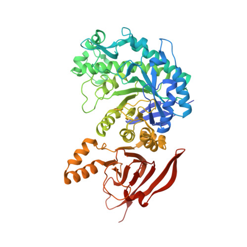
6TBI

Structure of a beta galactosidase with inhibitor


Experimental Data Snapshot

  • Method: X-RAY DIFFRACTION
  • Resolution: 1.46 Å
  • R-Value Free: 0.172 
  • R-Value Work: 0.126 
  • R-Value Observed: 0.128 

Starting Model: experimental
View more details

wwPDB Validation   3D Report Full Report


Ligand Structure Quality Assessment 


This is version 1.2 of the entry. See complete history


Literature

Mechanistic Insights into the Chaperoning of Human Lysosomal-Galactosidase Activity: Highly Functionalized Aminocyclopentanes and C -5a-Substituted Derivatives of 4- epi -Isofagomine.

Weber, P.Thonhofer, M.Averill, S.Davies, G.J.Santana, A.G.Muller, P.Nasseri, S.A.Offen, W.A.Pabst, B.M.Paschke, E.Schalli, M.Torvisco, A.Tschernutter, M.Tysoe, C.Windischhofer, W.Withers, S.G.Wolfsgruber, A.Wrodnigg, T.M.Stutz, A.E.

(2020) Molecules 25

  • DOI: https://doi.org/10.3390/molecules25174025
  • Primary Citation of Related Structures:  
    6TBF, 6TBG, 6TBH, 6TBI, 6TBJ, 6TBK

  • PubMed Abstract: 

    Glycosidase inhibitors have shown great potential as pharmacological chaperones for lysosomal storage diseases. In light of this, a series of new cyclopentanoid β-galactosidase inhibitors were prepared and their inhibitory and pharmacological chaperoning activities determined and compared with those of lipophilic analogs of the potent β-d-galactosidase inhibitor 4- epi -isofagomine. Structure-activity relationships were investigated by X-ray crystallography as well as by alterations in the cyclopentane moiety such as deoxygenation and replacement by fluorine of a "strategic" hydroxyl group. New compounds have revealed highly promising activities with a range of β-galactosidase-compromised human cell lines and may serve as leads towards new pharmacological chaperones for G M1 -gangliosidosis and Morquio B disease.


  • Organizational Affiliation

    Glycogroup, Institute of Chemistry and Technology of Biobased Systems, Graz University of Technology, Stremayrgasse 9, A-8010 Graz, Austria.


Macromolecules
Find similar proteins by:  (by identity cutoff)  |  3D Structure
Entity ID: 1
MoleculeChains Sequence LengthOrganismDetailsImage
Beta-galactosidase, putative, bgl35A
A, B, C, D, E
A, B, C, D, E, F, G, H
550Cellvibrio japonicusMutation(s): 0 
Gene Names: bgl35ACJA_2707
EC: 3.2.1.23
UniProt
Find proteins for B3PBE0 (Cellvibrio japonicus (strain Ueda107))
Explore B3PBE0 
Go to UniProtKB:  B3PBE0
Entity Groups  
Sequence Clusters30% Identity50% Identity70% Identity90% Identity95% Identity100% Identity
UniProt GroupB3PBE0
Sequence Annotations
Expand
  • Reference Sequence
Small Molecules
Ligands 3 Unique
IDChains Name / Formula / InChI Key2D Diagram3D Interactions
N8V (Subject of Investigation/LOI)
Query on N8V

Download Ideal Coordinates CCD File 
AA [auth C]
BB [auth F]
JA [auth D]
LB [auth G]
N [auth A]
AA [auth C],
BB [auth F],
JA [auth D],
LB [auth G],
N [auth A],
SB [auth H],
U [auth B],
UA [auth E]
(1S,2S,3S,6R)-4-(hydroxymethyl)-6-(octylamino)cyclohex-4-ene-1,2,3-triol
C15 H29 N O4
UPZUHYMBTUUPML-QPSCCSFWSA-N
ACT
Query on ACT

Download Ideal Coordinates CCD File 
EA [auth D]
EB [auth G]
FA [auth D]
FB [auth G]
GB [auth G]
EA [auth D],
EB [auth G],
FA [auth D],
FB [auth G],
GB [auth G],
HB [auth G],
J [auth A],
NA [auth E],
NB [auth H],
OA [auth E],
P [auth B],
PA [auth E],
Q [auth B],
QA [auth E],
X [auth C],
YA [auth F],
ZA [auth F]
ACETATE ION
C2 H3 O2
QTBSBXVTEAMEQO-UHFFFAOYSA-M
NA
Query on NA

Download Ideal Coordinates CCD File 
AB [auth F]
BA [auth D]
CA [auth D]
CB [auth G]
DA [auth D]
AB [auth F],
BA [auth D],
CA [auth D],
CB [auth G],
DA [auth D],
DB [auth G],
GA [auth D],
HA [auth D],
I [auth A],
IA [auth D],
IB [auth G],
JB [auth G],
K [auth A],
KA [auth E],
KB [auth G],
L [auth A],
LA [auth E],
M [auth A],
MA [auth E],
MB [auth H],
O [auth B],
OB [auth H],
PB [auth H],
QB [auth H],
R [auth B],
RA [auth E],
RB [auth H],
S [auth B],
SA [auth E],
T [auth B],
TA [auth E],
V [auth C],
VA [auth F],
W [auth C],
WA [auth F],
XA [auth F],
Y [auth C],
Z [auth C]
SODIUM ION
Na
FKNQFGJONOIPTF-UHFFFAOYSA-N
Experimental Data & Validation

Experimental Data

  • Method: X-RAY DIFFRACTION
  • Resolution: 1.46 Å
  • R-Value Free: 0.172 
  • R-Value Work: 0.126 
  • R-Value Observed: 0.128 
  • Space Group: P 1
Unit Cell:
Length ( Å )Angle ( ˚ )
a = 99.12α = 90.2
b = 115.91β = 89.96
c = 116.103γ = 90.08
Software Package:
Software NamePurpose
REFMACrefinement
PDB_EXTRACTdata extraction
DIALSdata reduction
Aimlessdata scaling
REFMACphasing

Structure Validation

View Full Validation Report


Ligand Structure Quality Assessment 


Entry History & Funding Information

Deposition Data


Funding OrganizationLocationGrant Number
Biotechnology and Biological Sciences Research CouncilUnited KingdomBB/R001162/1

Revision History  (Full details and data files)

  • Version 1.0: 2020-08-19
    Type: Initial release
  • Version 1.1: 2021-03-03
    Changes: Database references
  • Version 1.2: 2024-01-24
    Changes: Data collection, Database references, Refinement description