6T96 | pdb_00006t96

Photorhabdus laumondii subsp. laumondii lectin PLL3


Experimental Data Snapshot

  • Method: X-RAY DIFFRACTION
  • Resolution: 1.65 Å
  • R-Value Free: 
    0.196 (Depositor), 0.205 (DCC) 
  • R-Value Work: 
    0.169 (Depositor), 0.182 (DCC) 
  • R-Value Observed: 
    0.171 (Depositor) 

Starting Model: experimental
View more details

wwPDB Validation 3D Report Full Report

Validation slider image for 6T96

This is version 1.2 of the entry. See complete history

Literature

Lectin PLL3, a Novel Monomeric Member of the Seven-Bladed beta-Propeller Lectin Family.

Faltinek, L.Fujdiarova, E.Melicher, F.Houser, J.Kasakova, M.Kondakov, N.Kononov, L.Parkan, K.Vidal, S.Wimmerova, M.

(2019) Molecules 24

  • DOI: https://doi.org/10.3390/molecules24244540
  • Primary Citation Related Structures: 
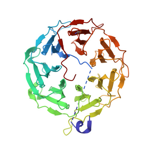
    6T96

  • PubMed Abstract: 

    The Photorhabdus species is a Gram-negative bacteria of the family Morganellaceae that is known for its mutualistic relationship with Heterorhabditis nematodes and pathogenicity toward insects. This study is focused on the characterization of the recombinant lectin PLL3 with an origin in P. laumondii subsp. laumondii . PLL3 belongs to the PLL family of lectins with a seven-bladed β-propeller fold. The binding properties of PLL3 were tested by hemagglutination assay, glycan array, isothermal titration calorimetry, and surface plasmon resonance, and its structure was determined by X-ray crystallography. Obtained data revealed that PLL3 binds similar carbohydrates to those that the other PLL family members bind, with some differences in the binding properties. PLL3 exhibited the highest affinity toward l-fucose and its derivatives but was also able to interact with O -methylated glycans and other ligands. Unlike the other members of this family, PLL3 was discovered to be a monomer, which might correspond to a weaker avidity effect compared to homologous lectins. Based on the similarity to the related lectins and their proposed biological function, PLL3 might accompany them during the interaction of P. laumondii with both the nematode partner and the insect host.


  • Organizational Affiliation
    • Department of Biochemistry, Faculty of Science, Masaryk University, Kotlářská 2, 611 37 Brno, Czech Republic.

Macromolecule Content 

  • Total Structure Weight: 40.55 kDa 
  • Atom Count: 3,114 
  • Modeled Residue Count: 348 
  • Deposited Residue Count: 371 
  • Unique protein chains: 1

Macromolecules

Find similar proteins by:|  3D Structure
Entity ID: 1
MoleculeChains  Sequence LengthOrganismDetailsImage
Lectin PLL3371Photorhabdus laumondii subsp. laumondii TTO1Mutation(s): 0 
Gene Names: plu0735
UniProt
Find proteins for Q7N8I7 (Photorhabdus laumondii subsp. laumondii (strain DSM 15139 / CIP 105565 / TT01))
Explore Q7N8I7 
Go to UniProtKB:  Q7N8I7
Entity Groups
Sequence Clusters30% Identity50% Identity70% Identity90% Identity95% Identity100% Identity
UniProt GroupQ7N8I7
Sequence Annotations
Expand
Reference Sequence

Small Molecules

Ligands 1 Unique
IDChains Name / Formula / InChI Key2D Diagram3D Interactions
NA

Query on NA


Download:Ideal Coordinates CCD File
B [auth A]SODIUM ION
Na
FKNQFGJONOIPTF-UHFFFAOYSA-N
Modified Residues  1 Unique
IDChains TypeFormula2D DiagramParent
CSO
Query on CSO
A
L-PEPTIDE LINKINGC3 H7 N O3 SCYS

Experimental Data & Validation

Experimental Data

  • Method: X-RAY DIFFRACTION
  • Resolution: 1.65 Å
  • R-Value Free:  0.196 (Depositor), 0.205 (DCC) 
  • R-Value Work:  0.169 (Depositor), 0.182 (DCC) 
  • R-Value Observed: 0.171 (Depositor) 
Space Group: P 21 21 21
Unit Cell:
Length ( Å )Angle ( ˚ )
a = 56.582α = 90
b = 69.416β = 90
c = 76.706γ = 90
Software Package:
Software NamePurpose
REFMACrefinement
SCALAdata scaling
PDB_EXTRACTdata extraction
XDSdata reduction
MOLREPphasing

Structure Validation

View Full Validation Report


Entry History 

Deposition Data

Revision History  (Full details and data files)

  • Version 1.0: 2019-12-25
    Type: Initial release
  • Version 1.1: 2024-01-24
    Changes: Data collection, Database references, Derived calculations, Refinement description
  • Version 1.2: 2024-11-20
    Changes: Structure summary