6T66 | pdb_00006t66

Crystal structure of the Vibrio cholerae replicative helicase (DnaB) with GDP-AlF4


Experimental Data Snapshot

  • Method: X-RAY DIFFRACTION
  • Resolution: 3.90 Å
  • R-Value Free: 
    0.276 (Depositor), 0.296 (DCC) 
  • R-Value Work: 
    0.255 (Depositor), 0.269 (DCC) 
  • R-Value Observed: 
    0.256 (Depositor) 

Starting Model: experimental
View more details

wwPDB Validation 3D Report Full Report

Validation slider image for 6T66

Ligand Structure Quality Assessment 


This is version 1.3 of the entry. See complete history

Literature

Study of the DnaB:DciA interplay reveals insights into the primary mode of loading of the bacterial replicative helicase.

Marsin, S.Adam, Y.Cargemel, C.Andreani, J.Baconnais, S.Legrand, P.Li de la Sierra-Gallay, I.Humbert, A.Aumont-Nicaise, M.Velours, C.Ochsenbein, F.Durand, D.Le Cam, E.Walbott, H.Possoz, C.Quevillon-Cheruel, S.Ferat, J.L.

(2021) Nucleic Acids Res 49: 6569-6586

  • DOI: https://doi.org/10.1093/nar/gkab463
  • Primary Citation Related Structures: 
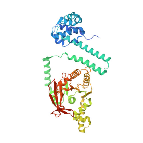
    6T66

  • PubMed Abstract: 

    Replicative helicases are essential proteins that unwind DNA in front of replication forks. Their loading depends on accessory proteins and in bacteria, DnaC and DnaI are well characterized loaders. However, most bacteria do not express either of these two proteins. Instead, they are proposed to rely on DciA, an ancestral protein unrelated to DnaC/I. While the DciA structure from Vibrio cholerae shares no homology with DnaC, it reveals similarities with DnaA and DnaX, two proteins involved during replication initiation. As other bacterial replicative helicases, VcDnaB adopts a toroid-shaped homo-hexameric structure, but with a slightly open dynamic conformation in the free state. We show that VcDnaB can load itself on DNA in vitro and that VcDciA stimulates this function, resulting in an increased DNA unwinding. VcDciA interacts with VcDnaB with a 3/6 stoichiometry and we show that a determinant residue, which discriminates DciA- and DnaC/I-helicases, is critical in vivo. Our work is the first step toward the understanding of the ancestral mode of loading of bacterial replicative helicases on DNA. It sheds light on the strategy employed by phage helicase loaders to hijack bacterial replicative helicases and may explain the recurrent domestication of dnaC/I through evolution in bacteria.


  • Organizational Affiliation
    • Université Paris-Saclay, CEA, CNRS, Institute for Integrative Biology of the Cell (I2BC), 91198, Gif-sur-Yvette, France.

Macromolecule Content 

  • Total Structure Weight: 321.05 kDa 
  • Atom Count: 20,822 
  • Modeled Residue Count: 2,644 
  • Deposited Residue Count: 2,844 
  • Unique protein chains: 1

Macromolecules

Find similar proteins by:|  3D Structure
Entity ID: 1
MoleculeChains  Sequence LengthOrganismDetailsImage
Replicative DNA helicase
A, B, C, D, E
A, B, C, D, E, F
474Vibrio choleraeMutation(s): 0 
Gene Names: dnaBBC353_11625C9J66_17780ERS013165_03687ERS013200_03939ERS013206_03692F0315_03560FLM02_14585HPY05_12185
EC: 3.6.4.12 (PDB Primary Data), 5.6.2.3 (UniProt)
UniProt
Find proteins for Q9KUY7 (Vibrio cholerae serotype O1 (strain ATCC 39315 / El Tor Inaba N16961))
Explore Q9KUY7 
Go to UniProtKB:  Q9KUY7
Entity Groups
Sequence Clusters30% Identity50% Identity70% Identity90% Identity95% Identity100% Identity
UniProt GroupQ9KUY7
Sequence Annotations
Expand
Reference Sequence

Small Molecules

Ligands 3 Unique
IDChains Name / Formula / InChI Key2D Diagram3D Interactions
GDP
(Subject of Investigation/LOI)

Query on GDP


Download:Ideal Coordinates CCD File
G [auth A]
J [auth B]
M [auth C]
P [auth D]
S [auth E]
G [auth A],
J [auth B],
M [auth C],
P [auth D],
S [auth E],
V [auth F]
GUANOSINE-5'-DIPHOSPHATE
C10 H15 N5 O11 P2
QGWNDRXFNXRZMB-UUOKFMHZSA-N
ALF
(Subject of Investigation/LOI)

Query on ALF


Download:Ideal Coordinates CCD File
I [auth A]
L [auth B]
O [auth C]
R [auth D]
U [auth E]
I [auth A],
L [auth B],
O [auth C],
R [auth D],
U [auth E],
X [auth F]
TETRAFLUOROALUMINATE ION
Al F4
UYOMQIYKOOHAMK-UHFFFAOYSA-J
MG
(Subject of Investigation/LOI)

Query on MG


Download:Ideal Coordinates CCD File
H [auth A]
K [auth B]
N [auth C]
Q [auth D]
T [auth E]
H [auth A],
K [auth B],
N [auth C],
Q [auth D],
T [auth E],
W [auth F]
MAGNESIUM ION
Mg
JLVVSXFLKOJNIY-UHFFFAOYSA-N

Experimental Data & Validation

Experimental Data

  • Method: X-RAY DIFFRACTION
  • Resolution: 3.90 Å
  • R-Value Free:  0.276 (Depositor), 0.296 (DCC) 
  • R-Value Work:  0.255 (Depositor), 0.269 (DCC) 
  • R-Value Observed: 0.256 (Depositor) 
Space Group: P 21 21 21
Unit Cell:
Length ( Å )Angle ( ˚ )
a = 118.978α = 90
b = 123.292β = 90
c = 263.493γ = 90
Software Package:
Software NamePurpose
BUSTERrefinement
XDSdata reduction
XDSdata scaling
MOLREPphasing

Structure Validation

View Full Validation Report


Ligand Structure Quality Assessment 


Entry History 

& Funding Information

Deposition Data


Funding OrganizationLocationGrant Number
French Infrastructure for Integrated Structural Biology (FRISBI)FranceANR-10-INBS-05

Revision History  (Full details and data files)

  • Version 1.0: 2021-04-28
    Type: Initial release
  • Version 1.1: 2021-06-23
    Changes: Database references
  • Version 1.2: 2021-06-30
    Changes: Database references
  • Version 1.3: 2024-01-24
    Changes: Data collection, Database references, Refinement description