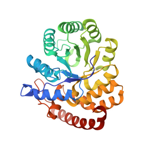
Structure of the 4-hydroxy-tetrahydrodipicolinate synthase from the thermoacidophilic methanotroph Methylacidiphilum fumariolicum SolV and the phylogeny of the aminotransferase pathway.
Schmitz, R.A., Dietl, A., Muller, M., Berben, T., Op den Camp, H.J.M., Barends, T.R.M.(2020) Acta Crystallogr F Struct Biol Commun 76: 199-208
- PubMed: 32356521 Search on PubMedSearch on PubMed Central
- DOI: https://doi.org/10.1107/S2053230X20005294
- Primary Citation Related Structures:
6T3T - PubMed Abstract:
The enzyme 4-hydroxy-tetrahydrodipicolinate synthase (DapA) is involved in the production of lysine and precursor molecules for peptidoglycan synthesis. In a multistep reaction, DapA converts pyruvate and L-aspartate-4-semialdehyde to 4-hydroxy-2,3,4,5-tetrahydrodipicolinic acid. In many organisms, lysine binds allosterically to DapA, causing negative feedback, thus making the enzyme an important regulatory component of the pathway. Here, the 2.1 Å resolution crystal structure of DapA from the thermoacidophilic methanotroph Methylacidiphilum fumariolicum SolV is reported. The enzyme crystallized as a contaminant of a protein preparation from native biomass. Genome analysis reveals that M. fumariolicum SolV utilizes the recently discovered aminotransferase pathway for lysine biosynthesis. Phylogenetic analyses of the genes involved in this pathway shed new light on the distribution of this pathway across the three domains of life.
- Department of Microbiology, Institute for Water and Wetland Research, Radboud University Nijmegen, 6525 AJ Nijmegen, The Netherlands.
Organizational Affiliation: