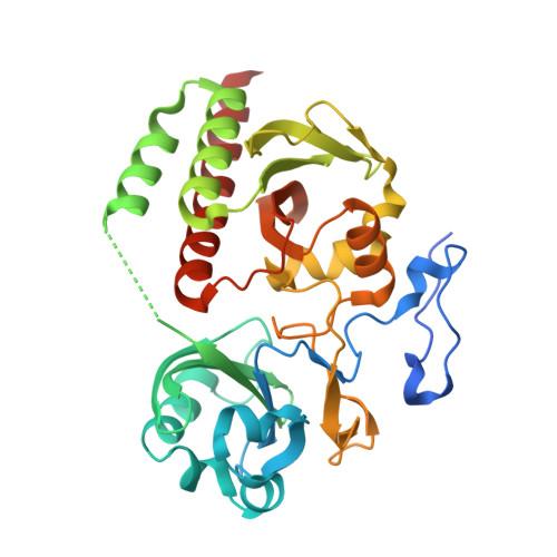
6T3L

PAS-GAF fragment from Deinococcus radiodurans phytochrome in dark state


Experimental Data Snapshot

  • Method: X-RAY DIFFRACTION
  • Resolution: 2.07 Å
  • R-Value Free: 0.192 
  • R-Value Work: 0.161 
  • R-Value Observed: 0.163 

Starting Model: experimental
View more details

wwPDB Validation   3D Report Full Report


Ligand Structure Quality Assessment 


This is version 1.3 of the entry. See complete history


Literature

The primary structural photoresponse of phytochrome proteins captured by a femtosecond X-ray laser.

Claesson, E.Wahlgren, W.Y.Takala, H.Pandey, S.Castillon, L.Kuznetsova, V.Henry, L.Panman, M.Carrillo, M.Kubel, J.Nanekar, R.Isaksson, L.Nimmrich, A.Cellini, A.Morozov, D.Maj, M.Kurttila, M.Bosman, R.Nango, E.Tanaka, R.Tanaka, T.Fangjia, L.Iwata, S.Owada, S.Moffat, K.Groenhof, G.A Stojkovic, E.A Ihalainen, J.Schmidt, M.Westenhoff, S.

(2020) Elife 9

  • DOI: https://doi.org/10.7554/eLife.53514
  • Primary Citation of Related Structures:  
    6T3L, 6T3U

  • PubMed Abstract: 

    Phytochrome proteins control the growth, reproduction, and photosynthesis of plants, fungi, and bacteria. Light is detected by a bilin cofactor, but it remains elusive how this leads to activation of the protein through structural changes. We present serial femtosecond X-ray crystallographic data of the chromophore-binding domains of a bacterial phytochrome at delay times of 1 ps and 10 ps after photoexcitation. The data reveal a twist of the D-ring, which leads to partial detachment of the chromophore from the protein. Unexpectedly, the conserved so-called pyrrole water is photodissociated from the chromophore, concomitant with movement of the A-ring and a key signaling aspartate. The changes are wired together by ultrafast backbone and water movements around the chromophore, channeling them into signal transduction towards the output domains. We suggest that the observed collective changes are important for the phytochrome photoresponse, explaining the earliest steps of how plants, fungi and bacteria sense red light.


  • Organizational Affiliation

    Department of Chemistry and Molecular Biology, University of Gothenburg, Gothenburg, Sweden.


Macromolecules
Find similar proteins by:  (by identity cutoff)  |  3D Structure
Entity ID: 1
MoleculeChains Sequence LengthOrganismDetailsImage
BacteriophytochromeA [auth B],
B [auth A]
343Deinococcus radioduransMutation(s): 0 
Gene Names: bphPDR_A0050
EC: 2.7.13.3
UniProt
Find proteins for Q9RZA4 (Deinococcus radiodurans (strain ATCC 13939 / DSM 20539 / JCM 16871 / CCUG 27074 / LMG 4051 / NBRC 15346 / NCIMB 9279 / VKM B-1422 / R1))
Explore Q9RZA4 
Go to UniProtKB:  Q9RZA4
Entity Groups  
Sequence Clusters30% Identity50% Identity70% Identity90% Identity95% Identity100% Identity
UniProt GroupQ9RZA4
Sequence Annotations
Expand
  • Reference Sequence
Small Molecules
Ligands 1 Unique
IDChains Name / Formula / InChI Key2D Diagram3D Interactions
LBV (Subject of Investigation/LOI)
Query on LBV

Download Ideal Coordinates CCD File 
C [auth B],
D [auth A]
3-[2-[(Z)-[3-(2-carboxyethyl)-5-[(Z)-(4-ethenyl-3-methyl-5-oxidanylidene-pyrrol-2-ylidene)methyl]-4-methyl-pyrrol-1-ium -2-ylidene]methyl]-5-[(Z)-[(3E)-3-ethylidene-4-methyl-5-oxidanylidene-pyrrolidin-2-ylidene]methyl]-4-methyl-1H-pyrrol-3- yl]propanoic acid
C33 H37 N4 O6
DKMLMZVDTGOEGU-ISEYCTJISA-O
Experimental Data & Validation

Experimental Data

Unit Cell:
Length ( Å )Angle ( ˚ )
a = 54.98α = 90
b = 116.69β = 90
c = 117.86γ = 90
Software Package:
Software NamePurpose
CrystFELdata reduction
CrystFELdata scaling
PHASERphasing
REFMACrefinement
PDB_EXTRACTdata extraction

Structure Validation

View Full Validation Report


Ligand Structure Quality Assessment 


Entry History & Funding Information

Deposition Data


Funding OrganizationLocationGrant Number
European Research CouncilSweden279944
Japan Agency for Medical Research and Development (AMED)Japan--
Academy of FinlandFinland285461
Academy of FinlandFinland296135

Revision History  (Full details and data files)

  • Version 1.0: 2020-04-08
    Type: Initial release
  • Version 1.1: 2023-12-13
    Changes: Data collection, Database references, Derived calculations, Structure summary
  • Version 1.2: 2024-01-24
    Changes: Refinement description
  • Version 1.3: 2024-11-13
    Changes: Structure summary