6T17

Cryo-EM structure of the wild-type flagellar filament of the Firmicute Kurthia


Experimental Data Snapshot

  • Method: ELECTRON MICROSCOPY
  • Resolution: 2.80 Å
  • Aggregation State: FILAMENT 
  • Reconstruction Method: HELICAL 

wwPDB Validation   3D Report Full Report


This is version 1.0 of the entry. See complete history


Literature

The wild-type flagellar filament of the Firmicute Kurthia at 2.8 angstrom resolution in vivo.

Blum, T.B.Filippidou, S.Fatton, M.Junier, P.Abrahams, J.P.

(2019) Sci Rep 9: 14948-14948

  • DOI: https://doi.org/10.1038/s41598-019-51440-1
  • Primary Citation of Related Structures:  
    6T17

  • PubMed Abstract: 

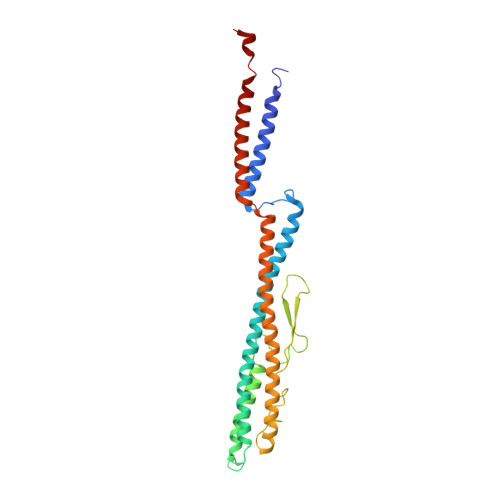
    Bacteria swim and swarm by rotating the micrometers long, helical filaments of their flagella. They change direction by reversing their flagellar rotation, which switches the handedness of the filament's supercoil. So far, all studied functional filaments are composed of a mixture of L- and R-state flagellin monomers. Here we show in a study of the wild type Firmicute Kurthia sp., that curved, functional filaments can adopt a conformation in vivo that is closely related to a uniform, all-L-state. This sheds additional light on transitions of the flagellar supercoil and uniquely reveals the atomic structure of a wild-type flagellar filament in vivo, including six residues showing clearly densities of O-linked glycosylation.


  • Organizational Affiliation

    Biology and Chemistry, Laboratory of Nanoscale Biology, Paul Scherrer Institute (PSI), CH-5232, Villigen, Switzerland. thorsten.blum@psi.ch.


Macromolecules
Find similar proteins by:  (by identity cutoff)  |  3D Structure
Entity ID: 1
MoleculeChains Sequence LengthOrganismDetailsImage
Flagellin276Kurthia sp. 11kri321Mutation(s): 0 
Gene Names: ASO14_2420
UniProt
Find proteins for A0A0X9VHV9 (Kurthia sp. 11kri321)
Explore A0A0X9VHV9 
Go to UniProtKB:  A0A0X9VHV9
Entity Groups  
Sequence Clusters30% Identity50% Identity70% Identity90% Identity95% Identity100% Identity
UniProt GroupA0A0X9VHV9
Sequence Annotations
Expand
  • Reference Sequence
Experimental Data & Validation

Experimental Data

  • Method: ELECTRON MICROSCOPY
  • Resolution: 2.80 Å
  • Aggregation State: FILAMENT 
  • Reconstruction Method: HELICAL 

Structure Validation

View Full Validation Report


Entry History & Funding Information

Deposition Data


Funding OrganizationLocationGrant Number
Swiss National Science FoundationSwitzerland31003A_17002
Swiss National Science FoundationSwitzerland200021_165669
Swiss National Science FoundationSwitzerland31003A_152972
Swiss National Science FoundationSwitzerland31003A_179297

Revision History  (Full details and data files)

  • Version 1.0: 2019-10-30
    Type: Initial release