6SY7 | pdb_00006sy7

Structure of Trypanosome Brucei Phosphofructokinase in complex with AMP.


Experimental Data Snapshot

  • Method: X-RAY DIFFRACTION
  • Resolution: 2.75 Å
  • R-Value Free: 
    0.227 (DCC) 
  • R-Value Work: 
    0.205 (DCC) 

Starting Model: experimental
View more details

wwPDB Validation 3D Report Full Report

Validation slider image for 6SY7

Ligand Structure Quality Assessment 


This is version 1.2 of the entry. See complete history

Literature

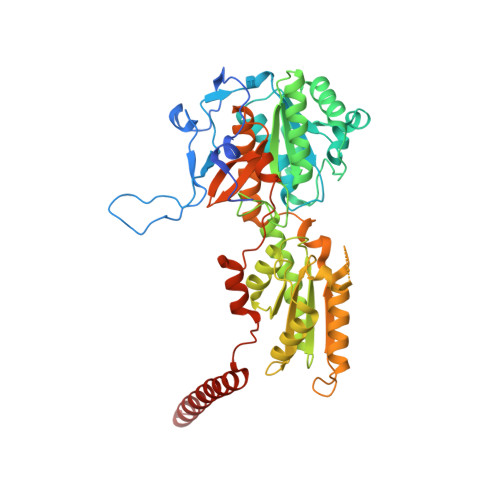
Kinetic and structural studies of Trypanosoma and Leishmania phosphofructokinases show evolutionary divergence and identify AMP as a switch regulating glycolysis versus gluconeogenesis.

Fernandes, P.M.Kinkead, J.McNae, I.W.Vasquez-Valdivieso, M.Wear, M.A.Michels, P.A.M.Walkinshaw, M.D.

(2020) FEBS J 287: 2847-2861

  • DOI: https://doi.org/10.1111/febs.15177
  • Primary Citation Related Structures: 
    6SY7

  • PubMed Abstract: 

    Trypanosomatids possess glycosome organelles that contain much of the glycolytic machinery, including phosphofructokinase (PFK). We present kinetic and structural data for PFK from three human pathogenic trypanosomatids, illustrating intriguing differences that may reflect evolutionary adaptations to differing ecological niches. The activity of Leishmania PFK - to a much larger extent than Trypanosoma PFK - is reliant on AMP for activity regulation, with 1 mm AMP increasing the L. infantum PFK (LiPFK) kcat/K 0.5 F6P value by 10-fold, compared to only a 1.3- and 1.4-fold increase for T. cruzi and T. brucei PFK, respectively. We also show that Leishmania PFK melts at a significantly lower (> 15 °C) temperature than Trypanosoma PFKs and that addition of either AMP or ATP results in a marked stabilization of the protein. Sequence comparisons of Trypanosoma spp. and Leishmania spp. show that divergence of the two genera involved amino acid substitutions that occur in the enzyme's 'reaching arms' and 'embracing arms' that determine tetramer stability. The dramatic effects of AMP on Leishmania activity compared with the Trypanosoma PFKs may be explained by differences between the T-to-R equilibria for the two families, with the low-melting Leishmania PFK favouring the flexible inactive T-state in the absence of AMP. Sequence comparisons along with the enzymatic and structural data presented here also suggest there was a loss of AMP-dependent regulation in Trypanosoma species rather than gain of this characteristic in Leishmania species and that AMP acts as a key regulator in Leishmania governing the balance between glycolysis and gluconeogenesis.


  • Organizational Affiliation
    • Centre for Translational and Chemical Biology, School of Biological Sciences, The University of Edinburgh, Edinburgh, UK.

Macromolecule Content 

  • Total Structure Weight: 432.89 kDa 
  • Atom Count: 28,937 
  • Modeled Residue Count: 3,618 
  • Deposited Residue Count: 3,896 
  • Unique protein chains: 1

Macromolecules

Find similar proteins by:|  3D Structure
Entity ID: 1
MoleculeChains  Sequence LengthOrganismDetailsImage
ATP-dependent 6-phosphofructokinase
A, B, C, D, E
A, B, C, D, E, F, G, H
487Trypanosoma brucei bruceiMutation(s): 0 
Gene Names: pfk
EC: 2.7.1.11
UniProt
Find proteins for O15648 (Trypanosoma brucei brucei)
Explore O15648 
Go to UniProtKB:  O15648
Entity Groups
Sequence Clusters30% Identity50% Identity70% Identity90% Identity95% Identity100% Identity
UniProt GroupO15648
Sequence Annotations
Expand
Reference Sequence

Small Molecules

Ligands 3 Unique
IDChains Name / Formula / InChI Key2D Diagram3D Interactions
AMP
(Subject of Investigation/LOI)

Query on AMP


Download:Ideal Coordinates CCD File
CA [auth G]
DA [auth H]
K [auth A]
L [auth B]
Q [auth C]
CA [auth G],
DA [auth H],
K [auth A],
L [auth B],
Q [auth C],
R [auth D],
W [auth F],
X [auth F]
ADENOSINE MONOPHOSPHATE
C10 H14 N5 O7 P
UDMBCSSLTHHNCD-KQYNXXCUSA-N
GOL

Query on GOL


Download:Ideal Coordinates CCD File
BA [auth G]
FA [auth H]
J [auth A]
N [auth B]
P [auth C]
BA [auth G],
FA [auth H],
J [auth A],
N [auth B],
P [auth C],
T [auth D],
V [auth E],
Z [auth F]
GLYCEROL
C3 H8 O3
PEDCQBHIVMGVHV-UHFFFAOYSA-N
BNZ

Query on BNZ


Download:Ideal Coordinates CCD File
AA [auth G]
EA [auth H]
I [auth A]
M [auth B]
O [auth C]
AA [auth G],
EA [auth H],
I [auth A],
M [auth B],
O [auth C],
S [auth D],
U [auth E],
Y [auth F]
BENZENE
C6 H6
UHOVQNZJYSORNB-UHFFFAOYSA-N

Experimental Data & Validation

Experimental Data

  • Method: X-RAY DIFFRACTION
  • Resolution: 2.75 Å
  • R-Value Free:  0.227 (DCC) 
  • R-Value Work:  0.205 (DCC) 
Space Group: P 21 21 21
Unit Cell:
Length ( Å )Angle ( ˚ )
a = 118.395α = 90
b = 133.711β = 90
c = 271.82γ = 90
Software Package:
Software NamePurpose
REFMACrefinement
xia2data reduction
xia2data scaling
PHASERphasing

Structure Validation

View Full Validation Report


Ligand Structure Quality Assessment 


Entry History 

Deposition Data

Revision History  (Full details and data files)

  • Version 1.0: 2019-12-25
    Type: Initial release
  • Version 1.1: 2020-07-15
    Changes: Database references
  • Version 1.2: 2024-01-24
    Changes: Data collection, Database references, Refinement description