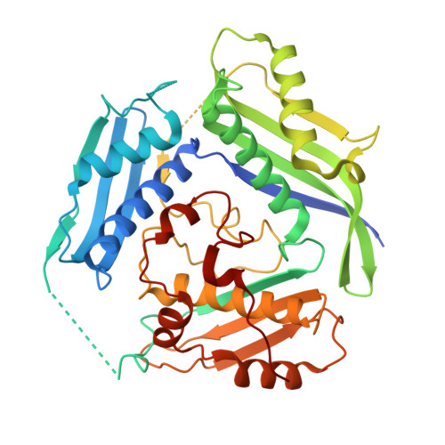
6SW5

Crystal structure of the human S-adenosylmethionine synthetase 1 (ligand-free form)


Experimental Data Snapshot

  • Method: X-RAY DIFFRACTION
  • Resolution: 2.35 Å
  • R-Value Free: 0.239 
  • R-Value Work: 0.188 
  • R-Value Observed: 0.191 

Starting Model: experimental
View more details

wwPDB Validation   3D Report Full Report


This is version 1.1 of the entry. See complete history


Literature

Structural basis of the dominant inheritance of hypermethioninemia associated with the Arg264His mutation in the MAT1A gene.

Panmanee, J.Antonyuk, S.V.Hasnain, S.S.

(2020) Acta Crystallogr D Struct Biol 76: 594-607

  • DOI: https://doi.org/10.1107/S2059798320006002
  • Primary Citation of Related Structures:  
    6SW5, 6SW6

  • PubMed Abstract: 

    Methionine adenosyltransferase (MAT) deficiency, characterized by isolated persistent hypermethioninemia (IPH), is caused by mutations in the MAT1A gene encoding MATαl, one of the major hepatic enzymes. Most of the associated hypermethioninemic conditions are inherited as autosomal recessive traits; however, dominant inheritance of hypermethioninemia is caused by an Arg264His (R264H) mutation. This mutation has been confirmed in a screening programme of newborns as the most common mutation in babies with IPH. Arg264 makes an inter-subunit salt bridge located at the dimer interface where the active site assembles. Here, it is demonstrated that the R264H mutation results in greatly reduced MAT activity, while retaining its ability to dimerize, indicating that the lower activity arises from alteration at the active site. The first crystallographic structure of the apo form of the wild-type MATαl enzyme is provided, which shows a tetrameric assembly in which two compact dimers combine to form a catalytic tetramer. In contrast, the crystal structure of the MATαl R264H mutant reveals a weaker dimeric assembly, suggesting that the mutation lowers the affinity for dimer-dimer interaction. The formation of a hetero-oligomer with the regulatory MATβV1 subunit or incubation with a quinolone-based compound (SCR0911) results in the near-full recovery of the enzymatic activity of the pathogenic mutation R264H, opening a clear avenue for a therapeutic solution based on chemical interventions that help to correct the defect of the enzyme in its ability to metabolize methionine.


  • Organizational Affiliation

    Molecular Biophysics Group, Institute of Systems, Molecular and Integrative Biology, Faculty of Health and Life Sciences, University of Liverpool, Crown Street, Liverpool L69 7ZB, United Kingdom.


Macromolecules
Find similar proteins by:  (by identity cutoff)  |  3D Structure
Entity ID: 1
MoleculeChains Sequence LengthOrganismDetailsImage
S-adenosylmethionine synthase isoform type-1
A, B, C, D
395Homo sapiensMutation(s): 0 
Gene Names: MAT1AAMS1MATA1
EC: 2.5.1.6
UniProt & NIH Common Fund Data Resources
Find proteins for Q00266 (Homo sapiens)
Explore Q00266 
Go to UniProtKB:  Q00266
PHAROS:  Q00266
GTEx:  ENSG00000151224 
Entity Groups  
Sequence Clusters30% Identity50% Identity70% Identity90% Identity95% Identity100% Identity
UniProt GroupQ00266
Sequence Annotations
Expand
  • Reference Sequence
Small Molecules
Ligands 2 Unique
IDChains Name / Formula / InChI Key2D Diagram3D Interactions
PEG
Query on PEG

Download Ideal Coordinates CCD File 
E [auth A],
L [auth B]
DI(HYDROXYETHYL)ETHER
C4 H10 O3
MTHSVFCYNBDYFN-UHFFFAOYSA-N
EDO
Query on EDO

Download Ideal Coordinates CCD File 
F [auth A]
G [auth A]
H [auth A]
I [auth A]
J [auth A]
F [auth A],
G [auth A],
H [auth A],
I [auth A],
J [auth A],
K [auth A],
M [auth B],
N [auth B],
O [auth C],
P [auth C],
Q [auth C],
R [auth D]
1,2-ETHANEDIOL
C2 H6 O2
LYCAIKOWRPUZTN-UHFFFAOYSA-N
Experimental Data & Validation

Experimental Data

  • Method: X-RAY DIFFRACTION
  • Resolution: 2.35 Å
  • R-Value Free: 0.239 
  • R-Value Work: 0.188 
  • R-Value Observed: 0.191 
  • Space Group: C 1 2 1
Unit Cell:
Length ( Å )Angle ( ˚ )
a = 218.684α = 90
b = 61β = 90.55
c = 119.762γ = 90
Software Package:
Software NamePurpose
Aimlessdata scaling
REFMACrefinement
PDB_EXTRACTdata extraction
xia2data reduction
MOLREPphasing

Structure Validation

View Full Validation Report


Entry History 

Deposition Data

Revision History  (Full details and data files)

  • Version 1.0: 2020-06-17
    Type: Initial release
  • Version 1.1: 2024-01-24
    Changes: Data collection, Database references, Refinement description