6STF | pdb_00006stf

Human Rab8a phosphorylated at Ser111 in complex with GDP


Experimental Data Snapshot

  • Method: X-RAY DIFFRACTION
  • Resolution: 2.40 Å
  • R-Value Free: 
    0.262 (Depositor), 0.270 (DCC) 
  • R-Value Work: 
    0.233 (Depositor), 0.250 (DCC) 
  • R-Value Observed: 
    0.234 (Depositor) 

Starting Model: experimental
View more details

wwPDB Validation   3D Report Full Report


Ligand Structure Quality Assessment 

Created with Raphaël 2.3.0Worse 01 BetterLigand structure goodness of fit to experimental dataBest fitted GDPClick on this verticalbar to view details

This is version 1.3 of the entry. See complete history


Literature

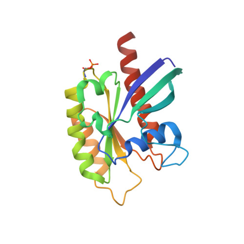
PINK1-dependent phosphorylation of Serine111 within the SF3 motif of Rab GTPases impairs effector interactions and LRRK2-mediated phosphorylation at Threonine72.

Vieweg, S.Mulholland, K.Brauning, B.Kachariya, N.Lai, Y.C.Toth, R.Singh, P.K.Volpi, I.Sattler, M.Groll, M.Itzen, A.Muqit, M.M.K.

(2020) Biochem J 477: 1651-1668

  • DOI: https://doi.org/10.1042/BCJ20190664
  • Primary Citation of Related Structures:  
    6STF, 6STG

  • PubMed Abstract: 

    Loss of function mutations in the PTEN-induced kinase 1 (PINK1) kinase are causal for autosomal recessive Parkinson's disease (PD) whilst gain of function mutations in the LRRK2 kinase cause autosomal dominant PD. PINK1 indirectly regulates the phosphorylation of a subset of Rab GTPases at a conserved Serine111 (Ser111) residue within the SF3 motif. Using genetic code expansion technologies, we have produced stoichiometric Ser111-phosphorylated Rab8A revealing impaired interactions with its cognate guanine nucleotide exchange factor and GTPase activating protein. In a screen for Rab8A kinases we identify TAK1 and MST3 kinases that can efficiently phosphorylate the Switch II residue Threonine72 (Thr72) in a similar manner as LRRK2 in vitro. Strikingly, we demonstrate that Ser111 phosphorylation negatively regulates the ability of LRRK2 but not MST3 or TAK1 to phosphorylate Thr72 of recombinant nucleotide-bound Rab8A in vitro and demonstrate an interplay of PINK1- and LRRK2-mediated phosphorylation of Rab8A in transfected HEK293 cells. Finally, we present the crystal structure of Ser111-phosphorylated Rab8A and nuclear magnetic resonance structure of Ser111-phosphorylated Rab1B. The structures reveal that the phosphorylated SF3 motif does not induce any major changes, but may interfere with effector-Switch II interactions through intramolecular H-bond formation and/or charge effects with Arg79. Overall, we demonstrate antagonistic regulation between PINK1-dependent Ser111 phosphorylation and LRRK2-mediated Thr72 phosphorylation of Rab8A indicating a potential cross-talk between PINK1-regulated mitochondrial homeostasis and LRRK2 signalling that requires further investigation in vivo.


  • Organizational Affiliation

    Department of Chemistry, Centre for Integrated Protein Science Munich (CIPSM), Technical University of Munich, Garching 85748, Germany.


Macromolecules
Find similar proteins by:  (by identity cutoff)  |  3D Structure
Entity ID: 1
MoleculeChains Sequence LengthOrganismDetailsImage
Ras-related protein Rab-8A
A, B, C, D, E
178Homo sapiensMutation(s): 0 
Gene Names: RAB8AMELRAB8
EC: 3.6.5.2
UniProt & NIH Common Fund Data Resources
Find proteins for P61006 (Homo sapiens)
Explore P61006 
Go to UniProtKB:  P61006
PHAROS:  P61006
GTEx:  ENSG00000167461 
Entity Groups  
Sequence Clusters30% Identity50% Identity70% Identity90% Identity95% Identity100% Identity
UniProt GroupP61006
Sequence Annotations
Expand
  • Reference Sequence
Small Molecules
Ligands 2 Unique
IDChains Name / Formula / InChI Key2D Diagram3D Interactions
GDP (Subject of Investigation/LOI)
Query on GDP

Download Ideal Coordinates CCD File 
F [auth A],
H [auth B],
J [auth C],
L [auth D],
N [auth E]
GUANOSINE-5'-DIPHOSPHATE
C10 H15 N5 O11 P2
QGWNDRXFNXRZMB-UUOKFMHZSA-N
MG (Subject of Investigation/LOI)
Query on MG

Download Ideal Coordinates CCD File 
G [auth A],
I [auth B],
K [auth C],
M [auth D],
O [auth E]
MAGNESIUM ION
Mg
JLVVSXFLKOJNIY-UHFFFAOYSA-N
Modified Residues  1 Unique
IDChains TypeFormula2D DiagramParent
SEP
Query on SEP
A, B, C, D, E
L-PEPTIDE LINKINGC3 H8 N O6 PSER
Experimental Data & Validation

Experimental Data

  • Method: X-RAY DIFFRACTION
  • Resolution: 2.40 Å
  • R-Value Free:  0.262 (Depositor), 0.270 (DCC) 
  • R-Value Work:  0.233 (Depositor), 0.250 (DCC) 
  • R-Value Observed: 0.234 (Depositor) 
Space Group: C 1 2 1
Unit Cell:
Length ( Å )Angle ( ˚ )
a = 117.55α = 90
b = 75.18β = 100.48
c = 107.7γ = 90
Software Package:
Software NamePurpose
REFMACrefinement
PDB_EXTRACTdata extraction
XDSdata reduction
XSCALEdata scaling
PHASERphasing

Structure Validation

View Full Validation Report


Ligand Structure Quality Assessment 

Created with Raphaël 2.3.0Worse 01 BetterLigand structure goodness of fit to experimental dataBest fitted GDPClick on this verticalbar to view details

Entry History & Funding Information

Deposition Data


Funding OrganizationLocationGrant Number
German Research FoundationGermanySFB1035

Revision History  (Full details and data files)

  • Version 1.0: 2020-04-22
    Type: Initial release
  • Version 1.1: 2020-05-20
    Changes: Database references
  • Version 1.2: 2024-01-24
    Changes: Data collection, Database references, Derived calculations, Refinement description
  • Version 1.3: 2024-10-16
    Changes: Structure summary