6SSX

cryo-em structure of alpha-synuclein fibril polymorph 2A


Experimental Data Snapshot

  • Method: ELECTRON MICROSCOPY
  • Resolution: 2.98 Å
  • Aggregation State: FILAMENT 
  • Reconstruction Method: HELICAL 

wwPDB Validation   3D Report Full Report


This is version 1.0 of the entry. See complete history


Literature

Two new polymorphic structures of human full-length alpha-synuclein fibrils solved by cryo-electron microscopy.

Guerrero-Ferreira, R.Taylor, N.M.Arteni, A.A.Kumari, P.Mona, D.Ringler, P.Britschgi, M.Lauer, M.E.Makky, A.Verasdonck, J.Riek, R.Melki, R.Meier, B.H.Bockmann, A.Bousset, L.Stahlberg, H.

(2019) Elife 8

  • DOI: https://doi.org/10.7554/eLife.48907
  • Primary Citation of Related Structures:  
    6RT0, 6RTB, 6SST, 6SSX

  • PubMed Abstract: 

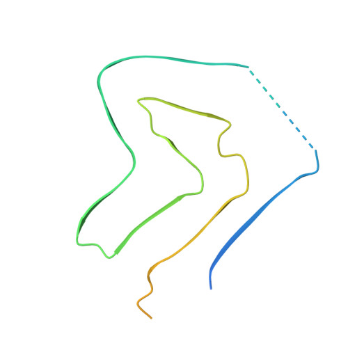
    Intracellular inclusions rich in alpha-synuclein are a hallmark of several neuropathological diseases including Parkinson's disease (PD). Previously, we reported the structure of alpha-synuclein fibrils (residues 1-121), composed of two protofibrils that are connected via a densely-packed interface formed by residues 50-57 (Guerrero-Ferreira, eLife 218;7:e36402). We here report two new polymorphic atomic structures of alpha-synuclein fibrils termed polymorphs 2a and 2b, at 3.0 Å and 3.4 Å resolution, respectively. These polymorphs show a radically different structure compared to previously reported polymorphs. The new structures have a 10 nm fibril diameter and are composed of two protofilaments which interact via intermolecular salt-bridges between amino acids K45, E57 (polymorph 2a) or E46 (polymorph 2b). The non-amyloid component (NAC) region of alpha-synuclein is fully buried by previously non-described interactions with the N-terminus. A hydrophobic cleft, the location of familial PD mutation sites, and the nature of the protofilament interface now invite to formulate hypotheses about fibril formation, growth and stability.


  • Organizational Affiliation

    Center for Cellular Imaging and NanoAnalytics (C-CINA), Biozentrum, University of Basel, Basel, Switzerland.


Macromolecules
Find similar proteins by:  (by identity cutoff)  |  3D Structure
Entity ID: 1
MoleculeChains Sequence LengthOrganismDetailsImage
Alpha-synuclein
A, B, C, D, E
A, B, C, D, E, F, G, H, I, J
140Homo sapiensMutation(s): 0 
Gene Names: SNCANACPPARK1
UniProt & NIH Common Fund Data Resources
Find proteins for P37840 (Homo sapiens)
Explore P37840 
Go to UniProtKB:  P37840
PHAROS:  P37840
GTEx:  ENSG00000145335 
Entity Groups  
Sequence Clusters30% Identity50% Identity70% Identity90% Identity95% Identity100% Identity
UniProt GroupP37840
Sequence Annotations
Expand
  • Reference Sequence
Experimental Data & Validation

Experimental Data

  • Method: ELECTRON MICROSCOPY
  • Resolution: 2.98 Å
  • Aggregation State: FILAMENT 
  • Reconstruction Method: HELICAL 
EM Software:
TaskSoftware PackageVersion
MODEL REFINEMENTPHENIX
RECONSTRUCTIONCoot

Structure Validation

View Full Validation Report


Entry History & Funding Information

Deposition Data


Funding OrganizationLocationGrant Number
Swiss National Science FoundationSwitzerlandCRSII3_154461
European UnionFrance116060
Novo Nordisk FoundationDenmarkNNF14CC0001
Swiss National Science FoundationSwitzerlandCRSII3_154461
Swiss National Science FoundationSwitzerlandCRSII5_177195
Swiss National Science FoundationSwitzerland20020_178792
French National Research AgencyFranceANR-12- BS08-0013-01
French National Research AgencyFranceANR-11-LABX-0048
French National Research AgencyFranceANR-11-IDEX-0007
Swiss National Science FoundationSwitzerland17.00038
French National Research AgencyFrench GuianaANR-17-JPCD-0002-02
French National Research AgencyFrench GuianaANR-17-JPCD-0005-01
French National Research AgencyFranceANR-10-INSB-05-01

Revision History  (Full details and data files)

  • Version 1.0: 2019-12-18
    Type: Initial release