6SSP | pdb_00006ssp

Structure of the pentameric ligand-gated ion channel ELIC in complex with a NAM nanobody


Experimental Data Snapshot

  • Method: X-RAY DIFFRACTION
  • Resolution: 3.25 Å
  • R-Value Free: 
    0.264 (Depositor), 0.268 (DCC) 
  • R-Value Work: 
    0.248 (Depositor), 0.263 (DCC) 
  • R-Value Observed: 
    0.249 (Depositor) 

Starting Models: experimental
View more details

wwPDB Validation 3D Report Full Report

Validation slider image for 6SSP

Ligand Structure Quality Assessment 


This is version 1.4 of the entry. See complete history

Literature

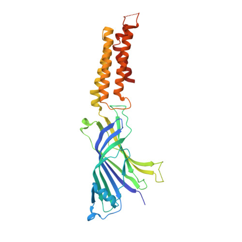
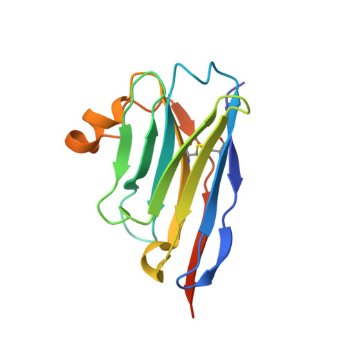
Modulation of the Erwinia ligand-gated ion channel (ELIC) and the 5-HT 3 receptor via a common vestibule site.

Brams, M.Govaerts, C.Kambara, K.Price, K.L.Spurny, R.Gharpure, A.Pardon, E.Evans, G.L.Bertrand, D.Lummis, S.C.Hibbs, R.E.Steyaert, J.Ulens, C.

(2020) Elife 9

  • DOI: https://doi.org/10.7554/eLife.51511
  • Primary Citation Related Structures: 
    6SSI, 6SSP

  • PubMed Abstract: 

    Pentameric ligand-gated ion channels (pLGICs) or Cys-loop receptors are involved in fast synaptic signaling in the nervous system. Allosteric modulators bind to sites that are remote from the neurotransmitter binding site, but modify coupling of ligand binding to channel opening. In this study, we developed nanobodies (single domain antibodies), which are functionally active as allosteric modulators, and solved co-crystal structures of the prokaryote ( Erwinia ) channel ELIC bound either to a positive or a negative allosteric modulator. The allosteric nanobody binding sites partially overlap with those of small molecule modulators, including a vestibule binding site that is not accessible in some pLGICs. Using mutagenesis, we extrapolate the functional importance of the vestibule binding site to the human 5-HT 3 receptor, suggesting a common mechanism of modulation in this protein and ELIC. Thus we identify key elements of allosteric binding sites, and extend drug design possibilities in pLGICs with an accessible vestibule site.


  • Organizational Affiliation
    • Laboratory of Structural Neurobiology, Department of Cellular and Molecular Medicine, Faculty of Medicine, KU Leuven, Leuven, Belgium.

Macromolecule Content 

  • Total Structure Weight: 400.44 kDa 
  • Atom Count: 26,239 
  • Modeled Residue Count: 3,260 
  • Deposited Residue Count: 3,458 
  • Unique protein chains: 2

Macromolecules

Find similar proteins by:|  3D Structure
Entity ID: 1
MoleculeChains  Sequence LengthOrganismDetailsImage
Cys-loop ligand-gated ion channel
A, B, C, D, E
A, B, C, D, E, F, G, H, I, J
318Dickeya chrysanthemiMutation(s): 0 
Membrane Entity: Yes 
UniProt
Find proteins for P0C7B7 (Dickeya chrysanthemi)
Explore P0C7B7 
Go to UniProtKB:  P0C7B7
Entity Groups
Sequence Clusters30% Identity50% Identity70% Identity90% Identity95% Identity100% Identity
UniProt GroupP0C7B7
Sequence Annotations
Expand
Reference Sequence
Find similar proteins by:|  3D Structure
Entity ID: 2
MoleculeChains  Sequence LengthOrganismDetailsImage
NANOBODY 21
K, L
139Lama glamaMutation(s): 0 

Small Molecules

Ligands 2 Unique
IDChains Name / Formula / InChI Key2D Diagram3D Interactions
UMQ

Query on UMQ


Download:Ideal Coordinates CCD File
AA [auth G]
CA [auth I]
DA [auth J]
N [auth A]
P [auth B]
AA [auth G],
CA [auth I],
DA [auth J],
N [auth A],
P [auth B],
Q [auth B],
S [auth C],
U [auth E],
X [auth F],
Z [auth G]
UNDECYL-MALTOSIDE
C23 H44 O11
UYEMNFYVTFDKRG-ZNGNCRBCSA-N
CA

Query on CA


Download:Ideal Coordinates CCD File
BA [auth H]
M [auth A]
O [auth B]
R [auth C]
T [auth D]
BA [auth H],
M [auth A],
O [auth B],
R [auth C],
T [auth D],
V [auth F],
W [auth F],
Y [auth G]
CALCIUM ION
Ca
BHPQYMZQTOCNFJ-UHFFFAOYSA-N

Experimental Data & Validation

Experimental Data

  • Method: X-RAY DIFFRACTION
  • Resolution: 3.25 Å
  • R-Value Free:  0.264 (Depositor), 0.268 (DCC) 
  • R-Value Work:  0.248 (Depositor), 0.263 (DCC) 
  • R-Value Observed: 0.249 (Depositor) 
Space Group: P 1
Unit Cell:
Length ( Å )Angle ( ˚ )
a = 121.804α = 71.02
b = 122.354β = 64.19
c = 128.221γ = 61.66
Software Package:
Software NamePurpose
XDSdata reduction
Aimlessdata scaling
MOLREPphasing
PHENIXrefinement
BUSTERrefinement
PDB_EXTRACTdata extraction

Structure Validation

View Full Validation Report


Ligand Structure Quality Assessment 


Entry History 

& Funding Information

Deposition Data


Funding OrganizationLocationGrant Number
Research Foundation - FlandersBelgium1200261
Research Foundation - FlandersBelgiumG.0762.13
KU LeuvenBelgiumOT/13/095
KU LeuvenBelgiumC32/16/035
KU LeuvenBelgiumC14/17/093

Revision History  (Full details and data files)

  • Version 1.0: 2020-02-12
    Type: Initial release
  • Version 1.1: 2020-02-19
    Changes: Database references
  • Version 1.2: 2020-09-30
    Changes: Database references, Derived calculations
  • Version 1.3: 2024-01-24
    Changes: Data collection, Database references, Refinement description
  • Version 1.4: 2024-10-23
    Changes: Structure summary