


Crystal structure of mouse PRMT6 in complex with inhibitors
Bonnefond, L., Cavarelli, J.To be published.
Experimental Data Snapshot
Starting Model: experimental
View more details
Entity ID: 1 | |||||
|---|---|---|---|---|---|
| Molecule | Chains | Sequence Length | Organism | Details | Image |
| Protein arginine N-methyltransferase 6 | 386 | Mus musculus | Mutation(s): 1 Gene Names: Prmt6, Hrmt1l6 EC: 2.1.1.319 | 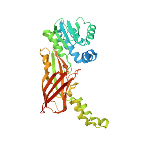 | |
UniProt & NIH Common Fund Data Resources | |||||
IMPC: MGI:2139971 | |||||
Entity Groups | |||||
| Sequence Clusters | 30% Identity50% Identity70% Identity90% Identity95% Identity100% Identity | ||||
| UniProt Group | Q6NZB1 | ||||
Sequence AnnotationsExpand | |||||
Reference Sequence | |||||
| Ligands 1 Unique | |||||
|---|---|---|---|---|---|
| ID | Chains | Name / Formula / InChI Key | 2D Diagram | 3D Interactions | |
| 6RE (Subject of Investigation/LOI) Download:Ideal Coordinates CCD File | C [auth A], D [auth B] | [[2-[(2~{R},3~{S},4~{R},5~{R})-5-(6-aminopurin-9-yl)-3,4-bis(oxidanyl)oxolan-2-yl]ethylamino]-azanyl-methylidene]azanium C12 H19 N8 O3 OIGRVZYOOMUALG-IOSLPCCCSA-O |  | ||
| Length ( Å ) | Angle ( ˚ ) |
|---|---|
| a = 41.526 | α = 90 |
| b = 117.979 | β = 103.3 |
| c = 71.945 | γ = 90 |
| Software Name | Purpose |
|---|---|
| PHENIX | refinement |
| PDB_EXTRACT | data extraction |
| XDS | data reduction |
| XDS | data scaling |
| PHASER | phasing |


