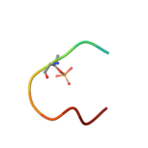
6SIQ

Fragment AZ-012 binding at the p53pT387/14-3-3 sigma interface


Experimental Data Snapshot

  • Method: X-RAY DIFFRACTION
  • Resolution: 1.60 Å
  • R-Value Free: 0.217 
  • R-Value Work: 0.182 

Starting Model: experimental
View more details

wwPDB Validation   3D Report Full Report


Ligand Structure Quality Assessment 


This is version 1.2 of the entry. See complete history


Literature

Fragment-based Differential Targeting of PPI Stabilizer Interfaces.

Guillory, X.Wolter, M.Leysen, S.Neves, J.F.Kuusk, A.Genet, S.Somsen, B.Morrow, J.K.Rivers, E.van Beek, L.Patel, J.Goodnow, R.Schoenherr, H.Fuller, N.Cao, Q.Doveston, R.G.Brunsveld, L.Arkin, M.R.Castaldi, P.Boyd, H.Landrieu, I.Chen, H.Ottmann, C.

(2020) J Med Chem 63: 6694-6707

  • DOI: https://doi.org/10.1021/acs.jmedchem.9b01942
  • Primary Citation of Related Structures:  
    6R5L, 6RHC, 6RJL, 6RJQ, 6RJZ, 6RK8, 6RKI, 6RKK, 6RKM, 6RL3, 6RL4, 6RL6, 6RM5, 6RM7, 6RP6, 6RWH, 6RWI, 6RWS, 6RWU, 6RX2, 6S39, 6S3C, 6S40, 6S9Q, 6SIN, 6SIO, 6SIP, 6SIQ, 6SLV, 6SLW, 6SLX

  • PubMed Abstract: 

    Stabilization of protein-protein interactions (PPIs) holds great potential for therapeutic agents, as illustrated by the successful drugs rapamycin and lenalidomide. However, how such interface-binding molecules can be created in a rational, bottom-up manner is a largely unanswered question. We report here how a fragment-based approach can be used to identify chemical starting points for the development of small-molecule stabilizers that differentiate between two different PPI interfaces of the adapter protein 14-3-3. The fragments discriminately bind to the interface of 14-3-3 with the recognition motif of either the tumor suppressor protein p53 or the oncogenic transcription factor TAZ. This X-ray crystallography driven study shows that the rim of the interface of individual 14-3-3 complexes can be targeted in a differential manner with fragments that represent promising starting points for the development of specific 14-3-3 PPI stabilizers.


  • Organizational Affiliation

    Laboratory of Chemical Biology, Department of Biomedical Engineering and Institute for Complex Molecular Systems, Eindhoven University of Technology, P.O. Box 513, 5600MB Eindhoven, The Netherlands.


Macromolecules
Find similar proteins by:  (by identity cutoff)  |  3D Structure
Entity ID: 1
MoleculeChains Sequence LengthOrganismDetailsImage
14-3-3 protein sigma236Homo sapiensMutation(s): 0 
Gene Names: SFNHME1
UniProt & NIH Common Fund Data Resources
Find proteins for P31947 (Homo sapiens)
Go to UniProtKB:  P31947
PHAROS:  P31947
GTEx:  ENSG00000175793 
Sequence Annotations
Expand
  • Reference Sequence

Find similar proteins by:  Sequence   |   3D Structure  

Entity ID: 2
MoleculeChains Sequence LengthOrganismDetailsImage
Cellular tumor antigen p53B [auth P]12Homo sapiensMutation(s): 0 
UniProt & NIH Common Fund Data Resources
Find proteins for P04637 (Homo sapiens)
Go to UniProtKB:  P04637
PHAROS:  P04637
GTEx:  ENSG00000141510 
Sequence Annotations
Expand
  • Reference Sequence
Small Molecules
Modified Residues  1 Unique
IDChains TypeFormula2D DiagramParent
TPO
Query on TPO
B [auth P]L-PEPTIDE LINKINGC4 H10 N O6 PTHR
Experimental Data & Validation

Experimental Data

  • Method: X-RAY DIFFRACTION
  • Resolution: 1.60 Å
  • R-Value Free: 0.217 
  • R-Value Work: 0.182 
  • Space Group: C 2 2 21
Unit Cell:
Length ( Å )Angle ( ˚ )
a = 82.34α = 90
b = 112.004β = 90
c = 62.572γ = 90
Software Package:
Software NamePurpose
PHENIXrefinement
MOSFLMdata reduction
Aimlessdata scaling

Structure Validation

View Full Validation Report


Ligand Structure Quality Assessment 


Entry History & Funding Information

Deposition Data


Funding OrganizationLocationGrant Number
European CommissionNetherlands--

Revision History  (Full details and data files)

  • Version 1.0: 2020-06-17
    Type: Initial release
  • Version 1.1: 2020-07-22
    Changes: Database references
  • Version 1.2: 2024-01-24
    Changes: Data collection, Database references, Refinement description