6SH7

Crystal structure of the human DEAH-helicase DHX15 in complex with the NKRF G-patch


Experimental Data Snapshot

  • Method: X-RAY DIFFRACTION
  • Resolution: 2.21 Å
  • R-Value Free: 0.251 
  • R-Value Work: 0.209 
  • R-Value Observed: 0.211 

wwPDB Validation   3D Report Full Report


This is version 1.1 of the entry. See complete history


Literature

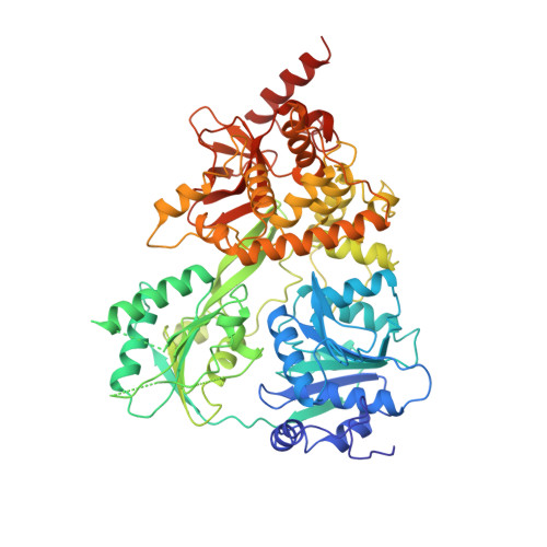
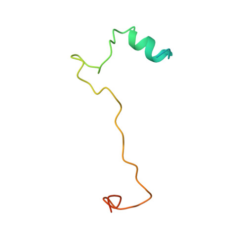
Structural basis for DEAH-helicase activation by G-patch proteins.

Studer, M.K.Ivanovic, L.Weber, M.E.Marti, S.Jonas, S.

(2020) Proc Natl Acad Sci U S A 117: 7159-7170

  • DOI: https://doi.org/10.1073/pnas.1913880117
  • Primary Citation of Related Structures:  
    6SH6, 6SH7

  • PubMed Abstract: 

    RNA helicases of the DEAH/RHA family are involved in many essential cellular processes, such as splicing or ribosome biogenesis, where they remodel large RNA-protein complexes to facilitate transitions to the next intermediate. DEAH helicases couple adenosine triphosphate (ATP) hydrolysis to conformational changes of their catalytic core. This movement results in translocation along RNA, which is held in place by auxiliary C-terminal domains. The activity of DEAH proteins is strongly enhanced by the large and diverse class of G-patch activators. Despite their central roles in RNA metabolism, insight into the molecular basis of G-patch-mediated helicase activation is missing. Here, we have solved the structure of human helicase DHX15/Prp43, which has a dual role in splicing and ribosome assembly, in complex with the G-patch motif of the ribosome biogenesis factor NKRF. The G-patch motif binds in an extended conformation across the helicase surface. It tethers the catalytic core to the flexibly attached C-terminal domains, thereby fixing a conformation that is compatible with RNA binding. Structures in the presence or absence of adenosine diphosphate (ADP) suggest that motions of the catalytic core, which are required for ATP binding, are still permitted. Concomitantly, RNA affinity, helicase, and ATPase activity of DHX15 are increased when G-patch is bound. Mutations that detach one end of the tether but maintain overall binding severely impair this enhancement. Collectively, our data suggest that the G-patch motif acts like a flexible brace between dynamic portions of DHX15 that restricts excessive domain motions but maintains sufficient flexibility for catalysis.


  • Organizational Affiliation

    Institute of Molecular Biology and Biophysics, Department of Biology, Swiss Federal Institute of Technology (ETH) Zürich, 8093 Zürich, Switzerland.


Macromolecules
Find similar proteins by:  (by identity cutoff)  |  3D Structure
Entity ID: 1
MoleculeChains Sequence LengthOrganismDetailsImage
Pre-mRNA-splicing factor ATP-dependent RNA helicase DHX15689Homo sapiensMutation(s): 0 
Gene Names: DHX15DBP1DDX15
EC: 3.6.4.13
UniProt & NIH Common Fund Data Resources
Find proteins for O43143 (Homo sapiens)
Explore O43143 
Go to UniProtKB:  O43143
PHAROS:  O43143
GTEx:  ENSG00000109606 
Entity Groups  
Sequence Clusters30% Identity50% Identity70% Identity90% Identity95% Identity100% Identity
UniProt GroupO43143
Sequence Annotations
Expand
  • Reference Sequence
Find similar proteins by:  (by identity cutoff)  |  3D Structure
Entity ID: 2
MoleculeChains Sequence LengthOrganismDetailsImage
NF-kappa-B-repressing factor67Homo sapiensMutation(s): 0 
Gene Names: NKRFITBA4NRF
UniProt & NIH Common Fund Data Resources
Find proteins for O15226 (Homo sapiens)
Explore O15226 
Go to UniProtKB:  O15226
PHAROS:  O15226
GTEx:  ENSG00000186416 
Entity Groups  
Sequence Clusters30% Identity50% Identity70% Identity90% Identity95% Identity100% Identity
UniProt GroupO15226
Sequence Annotations
Expand
  • Reference Sequence
Experimental Data & Validation

Experimental Data

  • Method: X-RAY DIFFRACTION
  • Resolution: 2.21 Å
  • R-Value Free: 0.251 
  • R-Value Work: 0.209 
  • R-Value Observed: 0.211 
  • Space Group: C 2 2 21
Unit Cell:
Length ( Å )Angle ( ˚ )
a = 82.243α = 90
b = 90.024β = 90
c = 213.885γ = 90
Software Package:
Software NamePurpose
PHENIXrefinement
XDSdata reduction
XSCALEdata scaling
PHASERphasing
Cootmodel building

Structure Validation

View Full Validation Report


Entry History & Funding Information

Deposition Data


Funding OrganizationLocationGrant Number
Swiss National Science FoundationSwitzerland31003A_179498
Swiss National Science FoundationSwitzerland182880

Revision History  (Full details and data files)

  • Version 1.0: 2020-04-22
    Type: Initial release
  • Version 1.1: 2024-01-24
    Changes: Data collection, Database references, Refinement description