6SH1

Crystal structure of substrate-free human neprilysin E584D.


Experimental Data Snapshot

  • Method: X-RAY DIFFRACTION
  • Resolution: 2.10 Å
  • R-Value Free: 0.261 
  • R-Value Work: 0.218 

wwPDB Validation   3D Report Full Report


Ligand Structure Quality Assessment 


This is version 1.3 of the entry. See complete history


Literature

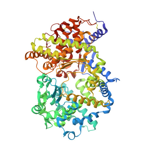
Crystal structure of peptide-bound neprilysin reveals key binding interactions.

Moss, S.Subramanian, V.Acharya, K.R.

(2020) FEBS Lett 594: 327-336

  • DOI: https://doi.org/10.1002/1873-3468.13602
  • Primary Citation of Related Structures:  
    6SH1, 6SH2

  • PubMed Abstract: 

    Neprilysin (NEP) is a promiscuous zinc metalloprotease with broad substrate specificity and cleaves a remarkable diversity of substrates through endopeptidase action. Two of these - amyloid-β and natriuretic peptides - implicate the enzyme in both Alzheimer's disease and cardiovascular disease, respectively. Here, we report the creation of a catalytically inactive NEP (E584D) to determine the first peptide-bound crystal structure at 2.6 Å resolution. The structure reveals key interactions involved in substrate binding which we have identified to be conserved in other known zinc metalloproteases. In addition, the structure provides evidence for a potential exosite within the central cavity that may play a critical role in substrate positioning. Together, these results contribute to our understanding of the molecular function of NEP.


  • Organizational Affiliation

    Department of Biology and Biochemistry, Claverton Down, University of Bath, UK.


Macromolecules
Find similar proteins by:  (by identity cutoff)  |  3D Structure
Entity ID: 1
MoleculeChains Sequence LengthOrganismDetailsImage
NeprilysinA [auth AAA],
B [auth CCC]
696Homo sapiensMutation(s): 0 
Gene Names: MMEEPN
EC: 3.4.24.11
UniProt & NIH Common Fund Data Resources
Find proteins for P08473 (Homo sapiens)
Explore P08473 
Go to UniProtKB:  P08473
PHAROS:  P08473
GTEx:  ENSG00000196549 
Entity Groups  
Sequence Clusters30% Identity50% Identity70% Identity90% Identity95% Identity100% Identity
UniProt GroupP08473
Sequence Annotations
Expand
  • Reference Sequence
Small Molecules
Ligands 3 Unique
IDChains Name / Formula / InChI Key2D Diagram3D Interactions
NAG
Query on NAG

Download Ideal Coordinates CCD File 
C [auth AAA]
D [auth AAA]
E [auth AAA]
F [auth AAA]
I [auth CCC]
C [auth AAA],
D [auth AAA],
E [auth AAA],
F [auth AAA],
I [auth CCC],
J [auth CCC],
K [auth CCC],
L [auth CCC]
2-acetamido-2-deoxy-beta-D-glucopyranose
C8 H15 N O6
OVRNDRQMDRJTHS-FMDGEEDCSA-N
ZN
Query on ZN

Download Ideal Coordinates CCD File 
H [auth AAA],
O [auth CCC]
ZINC ION
Zn
PTFCDOFLOPIGGS-UHFFFAOYSA-N
EDO
Query on EDO

Download Ideal Coordinates CCD File 
G [auth AAA],
M [auth CCC],
N [auth CCC]
1,2-ETHANEDIOL
C2 H6 O2
LYCAIKOWRPUZTN-UHFFFAOYSA-N
Experimental Data & Validation

Experimental Data

  • Method: X-RAY DIFFRACTION
  • Resolution: 2.10 Å
  • R-Value Free: 0.261 
  • R-Value Work: 0.218 
  • Space Group: P 1 21 1
Unit Cell:
Length ( Å )Angle ( ˚ )
a = 76.14α = 90
b = 99.745β = 106.231
c = 100.119γ = 90
Software Package:
Software NamePurpose
REFMACrefinement
DIALSdata reduction
Aimlessdata scaling
PHASERphasing

Structure Validation

View Full Validation Report


Ligand Structure Quality Assessment 


Entry History & Funding Information

Deposition Data


Funding OrganizationLocationGrant Number
Other privateUnited Kingdom286 (AS-PhD2015b006)

Revision History  (Full details and data files)

  • Version 1.0: 2019-09-25
    Type: Initial release
  • Version 1.1: 2020-02-05
    Changes: Data collection, Database references
  • Version 1.2: 2020-07-29
    Type: Remediation
    Reason: Carbohydrate remediation
    Changes: Data collection, Derived calculations, Structure summary
  • Version 1.3: 2024-01-24
    Changes: Data collection, Database references, Refinement description, Structure summary