6SG2 | pdb_00006sg2

FeFe Hydrogenase from Nitratidesulfovibrio vulgaris in Hinact state


Experimental Data Snapshot

  • Method: X-RAY DIFFRACTION
  • Resolution: 1.65 Å
  • R-Value Free: 
    0.212 (Depositor), 0.214 (DCC) 
  • R-Value Work: 
    0.171 (Depositor), 0.172 (DCC) 

Starting Model: experimental
View more details

wwPDB Validation 3D Report Full Report

Validation slider image for 6SG2

Ligand Structure Quality Assessment 


This is version 1.4 of the entry. See complete history

Literature

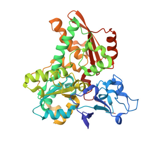
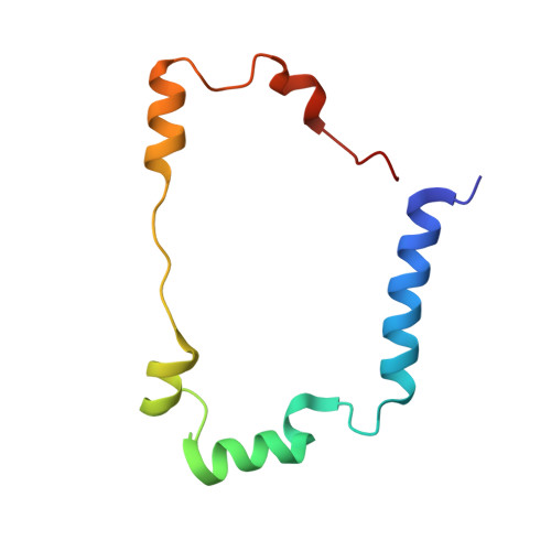
Caught in the H inact : Crystal Structure and Spectroscopy Reveal a Sulfur Bound to the Active Site of an O 2 -stable State of [FeFe] Hydrogenase.

Rodriguez-Macia, P.Galle, L.M.Bjornsson, R.Lorent, C.Zebger, I.Yoda, Y.Cramer, S.P.DeBeer, S.Span, I.Birrell, J.A.

(2020) Angew Chem Int Ed Engl 59: 16786-16794

  • DOI: https://doi.org/10.1002/anie.202005208
  • Primary Citation Related Structures: 
    6SG2

  • PubMed Abstract: 

    [FeFe] hydrogenases are the most active H 2 converting catalysts in nature, but their extreme oxygen sensitivity limits their use in technological applications. The [FeFe] hydrogenases from sulfate reducing bacteria can be purified in an O 2 -stable state called H inact . To date, the structure and mechanism of formation of H inact remain unknown. Our 1.65 Å crystal structure of this state reveals a sulfur ligand bound to the open coordination site. Furthermore, in-depth spectroscopic characterization by X-ray absorption spectroscopy (XAS), nuclear resonance vibrational spectroscopy (NRVS), resonance Raman (RR) spectroscopy and infrared (IR) spectroscopy, together with hybrid quantum mechanical and molecular mechanical (QM/MM) calculations, provide detailed chemical insight into the H inact state and its mechanism of formation. This may facilitate the design of O 2 -stable hydrogenases and molecular catalysts.


  • Organizational Affiliation
    • Department of Inorganic Spectroscopy, Max Planck Institute for Chemical Energy Conversion, Stiftstraße 34-36, 45470, Mülheim an der Ruhr, Germany.

Macromolecule Content 

  • Total Structure Weight: 54.75 kDa 
  • Atom Count: 3,936 
  • Modeled Residue Count: 484 
  • Deposited Residue Count: 484 
  • Unique protein chains: 2

Macromolecules

Find similar proteins by:|  3D Structure
Entity ID: 1
MoleculeChains  Sequence LengthOrganismDetailsImage
Periplasmic [Fe] hydrogenase large subunitA [auth AAA]396Nitratidesulfovibrio vulgarisMutation(s): 0 
Gene Names: hydADDE01_08480
Find similar proteins by:|  3D Structure
Entity ID: 2
MoleculeChains  Sequence LengthOrganismDetailsImage
HydBB [auth BBB]88Desulfovibrio desulfuricansMutation(s): 0 

Experimental Data & Validation

Experimental Data

  • Method: X-RAY DIFFRACTION
  • Resolution: 1.65 Å
  • R-Value Free:  0.212 (Depositor), 0.214 (DCC) 
  • R-Value Work:  0.171 (Depositor), 0.172 (DCC) 
Space Group: P 21 21 21
Unit Cell:
Length ( Å )Angle ( ˚ )
a = 49.34α = 90
b = 86.82β = 90
c = 88.17γ = 90
Software Package:
Software NamePurpose
REFMACrefinement
xia2data reduction
Aimlessdata scaling
PHASERphasing

Structure Validation

View Full Validation Report


Ligand Structure Quality Assessment 


Entry History 

& Funding Information

Deposition Data


Funding OrganizationLocationGrant Number
German Research FoundationGermanySP 1476/4-1

Revision History  (Full details and data files)

  • Version 1.0: 2020-07-08
    Type: Initial release
  • Version 1.1: 2020-09-30
    Changes: Database references
  • Version 1.2: 2020-12-16
    Changes: Database references
  • Version 1.3: 2024-01-24
    Changes: Data collection, Database references, Derived calculations, Refinement description
  • Version 1.4: 2026-02-25
    Changes: Source and taxonomy, Structure summary