6SF7

Atomic resolution structure of SplF protease from Staphylococcus aureus


Experimental Data Snapshot

  • Method: X-RAY DIFFRACTION
  • Resolution: 1.70 Å
  • R-Value Free: 0.208 
  • R-Value Work: 0.167 
  • R-Value Observed: 0.169 

wwPDB Validation   3D Report Full Report


This is version 1.2 of the entry. See complete history


Literature

Structural Determinants of Substrate Specificity of SplF Protease from Staphylococcus aureus .

Stach, N.Karim, A.Golik, P.Kitel, R.Pustelny, K.Gruba, N.Groborz, K.Jankowska, U.Kedracka-Krok, S.Wladyka, B.Drag, M.Lesner, A.Dubin, G.

(2021) Int J Mol Sci 22

  • DOI: https://doi.org/10.3390/ijms22042220
  • Primary Citation of Related Structures:  
    6SF7

  • PubMed Abstract: 

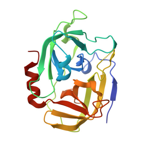
    Accumulating evidence suggests that six proteases encoded in the spl operon of a dangerous human pathogen, Staphylococcus aureus , may play a role in virulence. Interestingly, SplA, B, D, and E have complementary substrate specificities while SplF remains to be characterized in this regard. Here, we describe the prerequisites of a heterologous expression system for active SplF protease and characterize the enzyme in terms of substrate specificity and its structural determinants. Substrate specificity of SplF is comprehensively profiled using combinatorial libraries of peptide substrates demonstrating strict preference for long aliphatic sidechains at the P1 subsite and significant selectivity for aromatic residues at P3. The crystal structure of SplF was provided at 1.7 Å resolution to define the structural basis of substrate specificity of SplF. The obtained results were compared and contrasted with the characteristics of other Spl proteases determined to date to conclude that the spl operon encodes a unique extracellular proteolytic system.


  • Organizational Affiliation

    Malopolska Center of Biotechnology, Jagiellonian University, 7a Gronostajowa St., 30-387 Krakow, Poland.


Macromolecules
Find similar proteins by:  (by identity cutoff)  |  3D Structure
Entity ID: 1
MoleculeChains Sequence LengthOrganismDetailsImage
Serine protease SplF
A, B, C, D
205Staphylococcus aureus subsp. aureus USA300Mutation(s): 0 
Gene Names: splFSAUSA300_1753
EC: 3.4.21
UniProt
Find proteins for Q2FFT4 (Staphylococcus aureus (strain USA300))
Explore Q2FFT4 
Go to UniProtKB:  Q2FFT4
Entity Groups  
Sequence Clusters30% Identity50% Identity70% Identity90% Identity95% Identity100% Identity
UniProt GroupQ2FFT4
Sequence Annotations
Expand
  • Reference Sequence
Small Molecules
Ligands 4 Unique
IDChains Name / Formula / InChI Key2D Diagram3D Interactions
PG4
Query on PG4

Download Ideal Coordinates CCD File 
Q [auth C]TETRAETHYLENE GLYCOL
C8 H18 O5
UWHCKJMYHZGTIT-UHFFFAOYSA-N
PEG
Query on PEG

Download Ideal Coordinates CCD File 
E [auth A]DI(HYDROXYETHYL)ETHER
C4 H10 O3
MTHSVFCYNBDYFN-UHFFFAOYSA-N
SO4
Query on SO4

Download Ideal Coordinates CCD File 
F [auth A]
G [auth A]
H [auth A]
I [auth A]
J [auth B]
F [auth A],
G [auth A],
H [auth A],
I [auth A],
J [auth B],
K [auth B],
L [auth B],
M [auth B],
N [auth B],
P [auth C],
T [auth D],
U [auth D],
V [auth D],
W [auth D]
SULFATE ION
O4 S
QAOWNCQODCNURD-UHFFFAOYSA-L
EDO
Query on EDO

Download Ideal Coordinates CCD File 
O [auth B],
R [auth C],
S [auth C]
1,2-ETHANEDIOL
C2 H6 O2
LYCAIKOWRPUZTN-UHFFFAOYSA-N
Experimental Data & Validation

Experimental Data

  • Method: X-RAY DIFFRACTION
  • Resolution: 1.70 Å
  • R-Value Free: 0.208 
  • R-Value Work: 0.167 
  • R-Value Observed: 0.169 
  • Space Group: P 1
Unit Cell:
Length ( Å )Angle ( ˚ )
a = 56α = 104.57
b = 58.58β = 95.31
c = 62.3γ = 90.6
Software Package:
Software NamePurpose
REFMACrefinement
PDB_EXTRACTdata extraction
iMOSFLMdata reduction
SCALAdata scaling
PHASERphasing

Structure Validation

View Full Validation Report


Entry History 

Deposition Data

Revision History  (Full details and data files)

  • Version 1.0: 2021-03-03
    Type: Initial release
  • Version 1.1: 2021-03-24
    Changes: Database references
  • Version 1.2: 2024-05-15
    Changes: Data collection, Database references