6SE8 | pdb_00006se8

Cold-adapted beta-D-galactosidase from Arthrobacter sp. 32cB mutant E441Q


Experimental Data Snapshot

  • Method: X-RAY DIFFRACTION
  • Resolution: 1.83 Å
  • R-Value Free: 
    0.165 (Depositor), 0.172 (DCC) 
  • R-Value Work: 
    0.135 (Depositor), 0.146 (DCC) 
  • R-Value Observed: 
    0.135 (Depositor) 

Starting Model: experimental
View more details

wwPDB Validation 3D Report Full Report

Validation slider image for 6SE8

This is version 1.2 of the entry. See complete history

Literature

Active Site Architecture and Reaction Mechanism Determination of Cold Adapted beta-d-galactosidase fromArthrobactersp. 32cB.

Rutkiewicz, M.Bujacz, A.Wanarska, M.Wierzbicka-Wos, A.Cieslinski, H.

(2019) Int J Mol Sci 20

  • DOI: https://doi.org/10.3390/ijms20174301
  • Primary Citation Related Structures: 
    6SE8, 6SE9, 6SEA, 6SEB, 6SEC, 6SED

  • PubMed Abstract: 

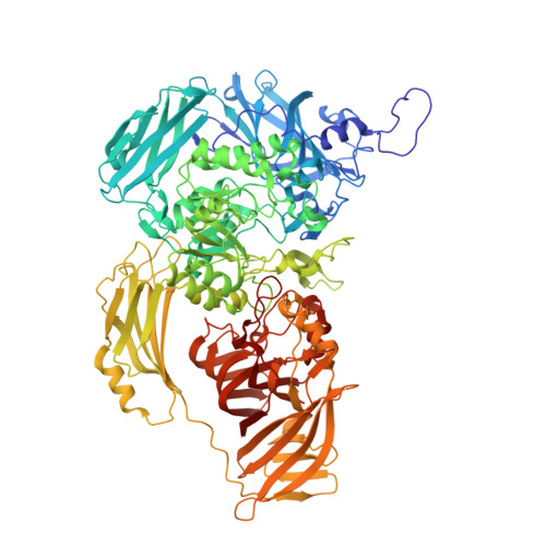
    Arth βDG is a dimeric, cold-adapted β-d-galactosidase that exhibits high hydrolytic and transglycosylation activity. A series of crystal structures of its wild form, as well as its Arth βDG_E441Q mutein complexes with ligands were obtained in order to describe the mode of its action. The Arth βDG_E441Q mutein is an inactive form of the enzyme designed to enable observation of enzyme interaction with its substrate. The resulting three-dimensional structures of complexes: Arth βDG_E441Q/LACs and Arth βDG/IPTG (ligand bound in shallow mode) and structures of complexes Arth βDG_E441Q/LACd, Arth βDG/ONPG (ligands bound in deep mode), and galactose Arth βDG/GAL and their analysis enabled structural characterization of the hydrolysis reaction mechanism. Furthermore, comparative analysis with mesophilic analogs revealed the most striking differences in catalysis mechanisms. The key role in substrate transfer from shallow to deep binding mode involves rotation of the F581 side chain. It is worth noting that the 10-aa loop restricting access to the active site in mesophilic GH2 βDGs, in Arth βDG is moved outward. This facilitates access of substrate to active site. Such a permanent exposure of the entrance to the active site may be a key factor for improved turnover rate of the cold adapted enzyme and thus a structural feature related to its cold adaptation.


  • Organizational Affiliation
    • Institute of Technical Biochemistry, Faculty of Biotechnology and Food Sciences, Lodz University of Technology, Stefanowskiego 4/10, 90-924 Lodz, Poland.

Macromolecule Content 

  • Total Structure Weight: 111.89 kDa 
  • Atom Count: 8,760 
  • Modeled Residue Count: 1,001 
  • Deposited Residue Count: 1,010 
  • Unique protein chains: 1

Macromolecules

Find similar proteins by:|  3D Structure
Entity ID: 1
MoleculeChains  Sequence LengthOrganismDetailsImage
Beta-galactosidase1,010Arthrobacter sp. 32cBMutation(s): 1 
EC: 3.2.1.23
UniProt
Find proteins for A0A023UGN9 (Arthrobacter sp. 32cB)
Explore A0A023UGN9 
Go to UniProtKB:  A0A023UGN9
Entity Groups
Sequence Clusters30% Identity50% Identity70% Identity90% Identity95% Identity100% Identity
UniProt GroupA0A023UGN9
Sequence Annotations
Expand
Reference Sequence

Small Molecules

Ligands 5 Unique
IDChains Name / Formula / InChI Key2D Diagram3D Interactions
EPE

Query on EPE


Download:Ideal Coordinates CCD File
EA [auth A]4-(2-HYDROXYETHYL)-1-PIPERAZINE ETHANESULFONIC ACID
C8 H18 N2 O4 S
JKMHFZQWWAIEOD-UHFFFAOYSA-N
MLI

Query on MLI


Download:Ideal Coordinates CCD File
AA [auth A]
BA [auth A]
CA [auth A]
DA [auth A]
X [auth A]
AA [auth A],
BA [auth A],
CA [auth A],
DA [auth A],
X [auth A],
Y [auth A],
Z [auth A]
MALONATE ION
C3 H2 O4
OFOBLEOULBTSOW-UHFFFAOYSA-L
ACT

Query on ACT


Download:Ideal Coordinates CCD File
B [auth A]
C [auth A]
D [auth A]
E [auth A]
F [auth A]
B [auth A],
C [auth A],
D [auth A],
E [auth A],
F [auth A],
G [auth A],
H [auth A],
I [auth A],
J [auth A],
K [auth A],
L [auth A],
M [auth A],
N [auth A],
O [auth A],
P [auth A],
Q [auth A]
ACETATE ION
C2 H3 O2
QTBSBXVTEAMEQO-UHFFFAOYSA-M
FMT

Query on FMT


Download:Ideal Coordinates CCD File
FA [auth A],
GA [auth A]
FORMIC ACID
C H2 O2
BDAGIHXWWSANSR-UHFFFAOYSA-N
NA

Query on NA


Download:Ideal Coordinates CCD File
R [auth A]
S [auth A]
T [auth A]
U [auth A]
V [auth A]
R [auth A],
S [auth A],
T [auth A],
U [auth A],
V [auth A],
W [auth A]
SODIUM ION
Na
FKNQFGJONOIPTF-UHFFFAOYSA-N

Experimental Data & Validation

Experimental Data

  • Method: X-RAY DIFFRACTION
  • Resolution: 1.83 Å
  • R-Value Free:  0.165 (Depositor), 0.172 (DCC) 
  • R-Value Work:  0.135 (Depositor), 0.146 (DCC) 
  • R-Value Observed: 0.135 (Depositor) 
Space Group: P 31 2 1
Unit Cell:
Length ( Å )Angle ( ˚ )
a = 136.859α = 90
b = 136.859β = 90
c = 127.082γ = 120
Software Package:
Software NamePurpose
XSCALEdata scaling
PHENIXrefinement
PDB_EXTRACTdata extraction
XDSdata reduction
PHENIXphasing

Structure Validation

View Full Validation Report


Entry History 

& Funding Information

Deposition Data


Funding OrganizationLocationGrant Number
Polish National Science CentrePoland2016/21/B/ST5/00555

Revision History  (Full details and data files)

  • Version 1.0: 2019-09-11
    Type: Initial release
  • Version 1.1: 2019-09-18
    Changes: Data collection, Database references
  • Version 1.2: 2024-01-24
    Changes: Data collection, Database references, Derived calculations, Refinement description