6SDZ | pdb_00006sdz

transthyritin derived amyloid fibril from patient with hereditary V30M ATTR amyloidosis


Experimental Data Snapshot

  • Method: ELECTRON MICROSCOPY
  • Resolution: 2.97 Å
  • Aggregation State: HELICAL ARRAY 
  • Reconstruction Method: HELICAL 

wwPDB Validation 3D Report Full Report

Validation slider image for 6SDZ

This is version 1.1 of the entry. See complete history

Literature

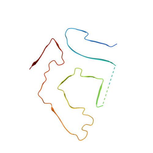
Cryo-EM structure of a transthyretin-derived amyloid fibril from a patient with hereditary ATTR amyloidosis.

Schmidt, M.Wiese, S.Adak, V.Engler, J.Agarwal, S.Fritz, G.Westermark, P.Zacharias, M.Fandrich, M.

(2019) Nat Commun 10: 5008-5008

  • DOI: https://doi.org/10.1038/s41467-019-13038-z
  • Primary Citation Related Structures: 
    6SDZ

  • PubMed Abstract: 

    ATTR amyloidosis is one of the worldwide most abundant forms of systemic amyloidosis. The disease is caused by the misfolding of transthyretin protein and the formation of amyloid deposits at different sites within the body. Here, we present a 2.97 Å cryo electron microscopy structure of a fibril purified from the tissue of a patient with hereditary Val30Met ATTR amyloidosis. The fibril consists of a single protofilament that is formed from an N-terminal and a C-terminal fragment of transthyretin. Our structure provides insights into the mechanism of misfolding and implies the formation of an early fibril state from unfolded transthyretin molecules, which upon proteolysis converts into mature ATTR amyloid fibrils.


  • Organizational Affiliation
    • Institute of Protein Biochemistry, Ulm University, 89081, Ulm, Germany.

Macromolecule Content 

  • Total Structure Weight: 151.9 kDa 
  • Atom Count: 7,909 
  • Modeled Residue Count: 1,012 
  • Deposited Residue Count: 1,397 
  • Unique protein chains: 1

Macromolecules

Find similar proteins by:|  3D Structure
Entity ID: 1
MoleculeChains  Sequence LengthOrganismDetailsImage
Transthyretin
A, B, C, D, E
A, B, C, D, E, F, G, H, I, J, K
127Homo sapiensMutation(s): 1 
UniProt & NIH Common Fund Data Resources
Find proteins for P02766 (Homo sapiens)
Explore P02766 
Go to UniProtKB:  P02766
PHAROS:  P02766
GTEx:  ENSG00000118271 
Entity Groups
Sequence Clusters30% Identity50% Identity70% Identity90% Identity95% Identity100% Identity
UniProt GroupP02766
Sequence Annotations
Expand
Reference Sequence

Experimental Data & Validation

Experimental Data

  • Method: ELECTRON MICROSCOPY
  • Resolution: 2.97 Å
  • Aggregation State: HELICAL ARRAY 
  • Reconstruction Method: HELICAL 
EM Software:
TaskSoftware PackageVersion
RECONSTRUCTIONRELION2.1
MODEL REFINEMENTPHENIX
MODEL REFINEMENTREFMAC

Structure Validation

View Full Validation Report


Entry History 

& Funding Information

Deposition Data


Funding OrganizationLocationGrant Number
German Research FoundationGermanySCHM 3276/1

Revision History  (Full details and data files)

  • Version 1.0: 2019-11-13
    Type: Initial release
  • Version 1.1: 2024-05-22
    Changes: Data collection, Database references