6SCZ

Mycobacterium tuberculosis alanine racemase inhibited by DCS


Experimental Data Snapshot

  • Method: X-RAY DIFFRACTION
  • Resolution: 1.57 Å
  • R-Value Free: 0.190 
  • R-Value Work: 0.150 
  • R-Value Observed: 0.152 

wwPDB Validation   3D Report Full Report


Ligand Structure Quality Assessment 


This is version 1.2 of the entry. See complete history


Literature

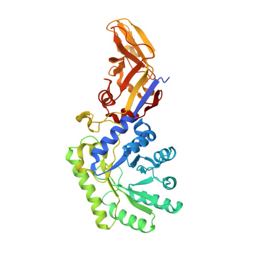
D-Cycloserine destruction by alanine racemase and the limit of irreversible inhibition.

de Chiara, C.Homsak, M.Prosser, G.A.Douglas, H.L.Garza-Garcia, A.Kelly, G.Purkiss, A.G.Tate, E.W.de Carvalho, L.P.S.

(2020) Nat Chem Biol 16: 686-694

  • DOI: https://doi.org/10.1038/s41589-020-0498-9
  • Primary Citation of Related Structures:  
    6SCZ

  • PubMed Abstract: 

    The broad-spectrum antibiotic D-cycloserine (DCS) is a key component of regimens used to treat multi- and extensively drug-resistant tuberculosis. DCS, a structural analog of D-alanine, binds to and inactivates two essential enzymes involved in peptidoglycan biosynthesis, alanine racemase (Alr) and D-Ala:D-Ala ligase. Inactivation of Alr is thought to proceed via a mechanism-based irreversible route, forming an adduct with the pyridoxal 5'-phosphate cofactor, leading to bacterial death. Inconsistent with this hypothesis, Mycobacterium tuberculosis Alr activity can be detected after exposure to clinically relevant DCS concentrations. To address this paradox, we investigated the chemical mechanism of Alr inhibition by DCS. Inhibition of M. tuberculosis Alr and other Alrs is reversible, mechanistically revealed by a previously unidentified DCS-adduct hydrolysis. Dissociation and subsequent rearrangement to a stable substituted oxime explains Alr reactivation in the cellular milieu. This knowledge provides a novel route for discovery of improved Alr inhibitors against M. tuberculosis and other bacteria.


  • Organizational Affiliation

    Mycobacterial Metabolism and Antibiotic Research Laboratory, The Francis Crick Institute, London, UK.


Macromolecules
Find similar proteins by:  (by identity cutoff)  |  3D Structure
Entity ID: 1
MoleculeChains Sequence LengthOrganismDetailsImage
Alanine racemase
A, B
387Mycobacterium tuberculosis H37RvMutation(s): 0 
Gene Names: alrRv3423cMTCY78.06
EC: 5.1.1.1
UniProt
Find proteins for P9WQA9 (Mycobacterium tuberculosis (strain ATCC 25618 / H37Rv))
Explore P9WQA9 
Go to UniProtKB:  P9WQA9
Entity Groups  
Sequence Clusters30% Identity50% Identity70% Identity90% Identity95% Identity100% Identity
UniProt GroupP9WQA9
Sequence Annotations
Expand
  • Reference Sequence
Small Molecules
Ligands 9 Unique
IDChains Name / Formula / InChI Key2D Diagram3D Interactions
P4K
Query on P4K

Download Ideal Coordinates CCD File 
VA [auth B],
WA [auth B],
XA [auth B],
Y [auth A],
Z [auth A]
polyethylene glycol
C30 H62 O15
WWPGFZAAWXFBTF-UHFFFAOYSA-N
OJQ
Query on OJQ

Download Ideal Coordinates CCD File 
BA [auth A],
YA [auth B]
[2-methyl-3-oxidanyl-5-(phosphonooxymethyl)pyridin-4-yl]methyl-(3-oxidanyl-1,2-oxazol-4-yl)azanium
C11 H15 N3 O7 P
PXWFNGNWQUPGPJ-UHFFFAOYSA-O
L7N (Subject of Investigation/LOI)
Query on L7N

Download Ideal Coordinates CCD File 
AA [auth A](~{E})-[2-methyl-3-oxidanyl-5-(phosphonooxymethyl)pyridin-4-yl]methylidene-[(4~{R})-3-oxidanylidene-1,2-oxazolidin-4-yl]azanium
C11 H15 N3 O7 P
PWALIMMCDFLLIJ-RZTFRGLUSA-O
TRS
Query on TRS

Download Ideal Coordinates CCD File 
CA [auth A],
ZA [auth B]
2-AMINO-2-HYDROXYMETHYL-PROPANE-1,3-DIOL
C4 H12 N O3
LENZDBCJOHFCAS-UHFFFAOYSA-O
PEG
Query on PEG

Download Ideal Coordinates CCD File 
SA [auth B]
TA [auth B]
U [auth A]
UA [auth B]
V [auth A]
SA [auth B],
TA [auth B],
U [auth A],
UA [auth B],
V [auth A],
W [auth A],
X [auth A]
DI(HYDROXYETHYL)ETHER
C4 H10 O3
MTHSVFCYNBDYFN-UHFFFAOYSA-N
GOL
Query on GOL

Download Ideal Coordinates CCD File 
C [auth A]
D [auth A]
DA [auth B]
E [auth A]
EA [auth B]
C [auth A],
D [auth A],
DA [auth B],
E [auth A],
EA [auth B],
F [auth A],
FA [auth B]
GLYCEROL
C3 H8 O3
PEDCQBHIVMGVHV-UHFFFAOYSA-N
EDO
Query on EDO

Download Ideal Coordinates CCD File 
HA [auth B]
IA [auth B]
J [auth A]
JA [auth B]
K [auth A]
HA [auth B],
IA [auth B],
J [auth A],
JA [auth B],
K [auth A],
KA [auth B],
L [auth A],
LA [auth B],
M [auth A],
MA [auth B],
N [auth A],
NA [auth B],
O [auth A],
OA [auth B],
P [auth A],
PA [auth B],
Q [auth A],
QA [auth B],
R [auth A],
RA [auth B],
S [auth A],
T [auth A]
1,2-ETHANEDIOL
C2 H6 O2
LYCAIKOWRPUZTN-UHFFFAOYSA-N
CA
Query on CA

Download Ideal Coordinates CCD File 
G [auth A]CALCIUM ION
Ca
BHPQYMZQTOCNFJ-UHFFFAOYSA-N
CL
Query on CL

Download Ideal Coordinates CCD File 
GA [auth B],
H [auth A],
I [auth A]
CHLORIDE ION
Cl
VEXZGXHMUGYJMC-UHFFFAOYSA-M
Experimental Data & Validation

Experimental Data

  • Method: X-RAY DIFFRACTION
  • Resolution: 1.57 Å
  • R-Value Free: 0.190 
  • R-Value Work: 0.150 
  • R-Value Observed: 0.152 
  • Space Group: P 41 21 2
Unit Cell:
Length ( Å )Angle ( ˚ )
a = 164.178α = 90
b = 164.178β = 90
c = 57.742γ = 90
Software Package:
Software NamePurpose
PHENIXrefinement
DIALSdata reduction
DIALSdata scaling
PHASERphasing

Structure Validation

View Full Validation Report


Ligand Structure Quality Assessment 


Entry History & Funding Information

Deposition Data


Funding OrganizationLocationGrant Number
The Francis Crick InstituteUnited KingdomSee list below
Cancer Research UKUnited KingdomFC001060
Medical Research Council (MRC, United Kingdom)United KingdomFC001060
Wellcome TrustUnited KingdomFC001060
Wellcome TrustUnited Kingdom104785/B/14/Z

Revision History  (Full details and data files)

  • Version 1.0: 2020-04-01
    Type: Initial release
  • Version 1.1: 2020-06-03
    Changes: Database references
  • Version 1.2: 2024-01-24
    Changes: Data collection, Database references, Derived calculations, Refinement description