6SCI | pdb_00006sci

Structure of AdhE form 1


Experimental Data Snapshot

  • Method: X-RAY DIFFRACTION
  • Resolution: 1.95 Å
  • R-Value Free: 
    0.217 (Depositor), 0.224 (DCC) 
  • R-Value Work: 
    0.182 (Depositor), 0.190 (DCC) 
  • R-Value Observed: 
    0.184 (Depositor) 

Starting Model: experimental
View more details

wwPDB Validation 3D Report Full Report

Validation slider image for 6SCI

This is version 2.1 of the entry. See complete history

Literature

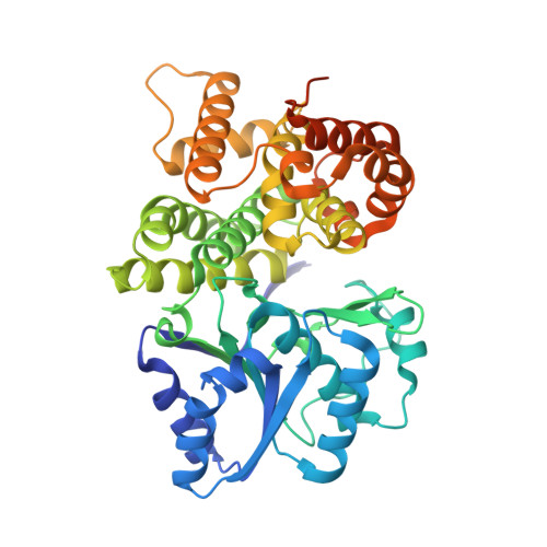
High-resolution structure of the alcohol dehydrogenase domain of the bifunctional bacterial enzyme AdhE.

Azmi, L.Bragginton, E.C.Cadby, I.T.Byron, O.Roe, A.J.Lovering, A.L.Gabrielsen, M.

(2020) Acta Crystallogr F Struct Biol Commun 76: 414-421

  • DOI: https://doi.org/10.1107/S2053230X20010237
  • Primary Citation Related Structures: 
    6SCG, 6SCI

  • PubMed Abstract: 

    The bifunctional alcohol/aldehyde dehydrogenase (AdhE) comprises both an N-terminal aldehyde dehydrogenase (AldDH) and a C-terminal alcohol dehydrogenase (ADH). In vivo, full-length AdhE oligomerizes into long oligomers known as spirosomes. However, structural analysis of AdhE is challenging owing to the heterogeneity of the spirosomes. Therefore, the domains of AdhE are best characterized separately. Here, the structure of ADH from the pathogenic Escherichia coli O157:H7 was determined to 1.65 Å resolution. The dimeric crystal structure was confirmed in solution by small-angle X-ray scattering.


  • Organizational Affiliation
    • Institute of Infection, Immunity and Inflammation, University of Glasgow, University Avenue, Glasgow G12 8QQ, United Kingdom.

Macromolecule Content 

  • Total Structure Weight: 99.9 kDa 
  • Atom Count: 6,351 
  • Modeled Residue Count: 806 
  • Deposited Residue Count: 910 
  • Unique protein chains: 1

Macromolecules

Find similar proteins by:|  3D Structure
Entity ID: 1
MoleculeChains  Sequence LengthOrganismDetailsImage
Aldehyde-alcohol dehydrogenase
A, B
455Escherichia coli K-12Mutation(s): 0 
Gene Names: adhEanab1241JW1228
EC: 1.1.1.1 (PDB Primary Data), 1.2.1.10 (PDB Primary Data)
UniProt
Find proteins for P0A9Q7 (Escherichia coli (strain K12))
Explore P0A9Q7 
Go to UniProtKB:  P0A9Q7
Entity Groups
Sequence Clusters30% Identity50% Identity70% Identity90% Identity95% Identity100% Identity
UniProt GroupP0A9Q7
Sequence Annotations
Expand
Reference Sequence

Experimental Data & Validation

Experimental Data

  • Method: X-RAY DIFFRACTION
  • Resolution: 1.95 Å
  • R-Value Free:  0.217 (Depositor), 0.224 (DCC) 
  • R-Value Work:  0.182 (Depositor), 0.190 (DCC) 
  • R-Value Observed: 0.184 (Depositor) 
Space Group: P 21 21 21
Unit Cell:
Length ( Å )Angle ( ˚ )
a = 71.03α = 90
b = 96.73β = 90
c = 122.89γ = 90
Software Package:
Software NamePurpose
REFMACrefinement
PHENIXrefinement
XDSdata reduction
Aimlessdata scaling
PHASERphasing
PDB_EXTRACTdata extraction

Structure Validation

View Full Validation Report


Entry History 

Deposition Data

Revision History  (Full details and data files)

  • Version 1.0: 2020-08-26
    Type: Initial release
  • Version 2.0: 2021-03-17
    Changes: Advisory, Atomic model, Data collection, Database references, Derived calculations, Experimental preparation, Polymer sequence, Refinement description, Source and taxonomy, Structure summary
  • Version 2.1: 2024-01-24
    Changes: Data collection, Database references, Refinement description