6S6Y | pdb_00006s6y

X-ray crystal structure of the formyltransferase/hydrolase complex (FhcABCD) from Methylorubrum extorquens in complex with methylofuran


Experimental Data Snapshot

  • Method: X-RAY DIFFRACTION
  • Resolution: 3.10 Å
  • R-Value Free: 
    0.251 (Depositor), 0.223 (DCC) 
  • R-Value Work: 
    0.221 (Depositor), 0.224 (DCC) 
  • R-Value Observed: 
    0.223 (Depositor) 

Starting Model: experimental
View more details

wwPDB Validation 3D Report Full Report

Validation slider image for 6S6Y

Ligand Structure Quality Assessment 


This is version 1.3 of the entry. See complete history

Literature

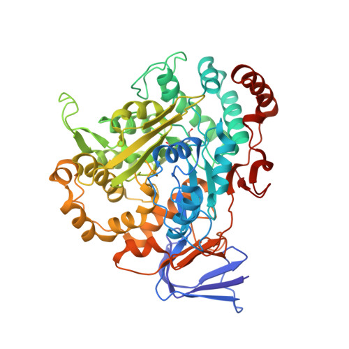
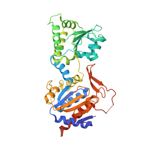
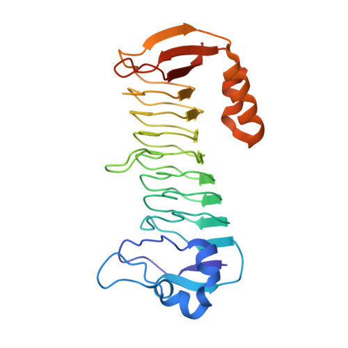
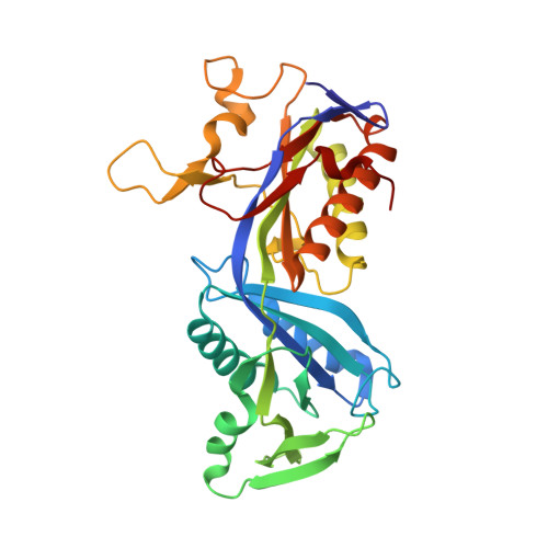
Methylofuran is a prosthetic group of the formyltransferase/hydrolase complex and shuttles one-carbon units between two active sites.

Hemmann, J.L.Wagner, T.Shima, S.Vorholt, J.A.

(2019) Proc Natl Acad Sci U S A 116: 25583-25590

  • DOI: https://doi.org/10.1073/pnas.1911595116
  • Primary Citation Related Structures: 
    6S6Y

  • PubMed Abstract: 

    Methylotrophy, the ability of microorganisms to grow on reduced one-carbon substrates such as methane or methanol, is a feature of various bacterial species. The prevailing oxidation pathway depends on tetrahydromethanopterin (H 4 MPT) and methylofuran (MYFR), an analog of methanofuran from methanogenic archaea. Formyltransferase/hydrolase complex (Fhc) generates formate from formyl-H 4 MPT in two consecutive reactions where MYFR acts as a carrier of one-carbon units. Recently, we chemically characterized MYFR from the model methylotroph Methylorubrum extorquens and identified an unusually long polyglutamate side chain of up to 24 glutamates. Here, we report on the crystal structure of Fhc to investigate the function of the polyglutamate side chain in MYFR and the relatedness of the enzyme complex with the orthologous enzymes in archaea. We identified MYFR as a prosthetic group that is tightly, but noncovalently, bound to Fhc. Surprisingly, the structure of Fhc together with MYFR revealed that the polyglutamate side chain of MYFR is branched and contains glutamates with amide bonds at both their α- and γ-carboxyl groups. This negatively charged and branched polyglutamate side chain interacts with a cluster of conserved positively charged residues of Fhc, allowing for strong interactions. The MYFR binding site is located equidistantly from the active site of the formyltransferase (FhcD) and metallo-hydrolase (FhcA). The polyglutamate serves therefore an additional function as a swinging linker to shuttle the one-carbon carrying amine between the two active sites, thereby likely increasing overall catalysis while decreasing the need for high intracellular MYFR concentrations.


  • Organizational Affiliation
    • Institute of Microbiology, Eidgenössische Technische Hochschule Zurich, 8093 Zurich, Switzerland.

Macromolecule Content 

  • Total Structure Weight: 628.8 kDa 
  • Atom Count: 43,288 
  • Modeled Residue Count: 5,894 
  • Deposited Residue Count: 5,980 
  • Unique protein chains: 4

Macromolecules

Find similar proteins by:|  3D Structure
Entity ID: 1
MoleculeChains  Sequence LengthOrganismDetailsImage
Formylmethanofuran dehydrogenase subunit A
A, E, I, M
548Methylorubrum extorquens PA1Mutation(s): 0 
Find similar proteins by:|  3D Structure
Entity ID: 2
MoleculeChains  Sequence LengthOrganismDetailsImage
Tungsten-containing formylmethanofuran dehydrogenase, subunit B
B, F, J, N
361Methylorubrum extorquens PA1Mutation(s): 0 
Find similar proteins by:|  3D Structure
Entity ID: 3
MoleculeChains  Sequence LengthOrganismDetailsImage
Formylmethanofuran dehydrogenase subunit C
C, G, K, O
276Methylorubrum extorquens PA1Mutation(s): 0 
Gene Names: Mext_1824
Find similar proteins by:|  3D Structure
Entity ID: 4
MoleculeChains  Sequence LengthOrganismDetailsImage
Formylmethanofuran--tetrahydromethanopterin formyltransferase
D, H, L, P
310Methylorubrum extorquens PA1Mutation(s): 0 
EC: 2.3.1.101

Small Molecules

Ligands 14 Unique
IDChains Name / Formula / InChI Key2D Diagram3D Interactions
MFN
(Subject of Investigation/LOI)

Query on MFN


Download:Ideal Coordinates CCD File
R [auth A]N-[4,5,7-TRICARBOXYHEPTANOYL]-L-GAMMA-GLUTAMYL-N-{2-[4-({5-[(FORMYLAMINO)METHYL]-3-FURYL}METHOXY)PHENYL]ETHYL}-D-GLUTAMINE
C35 H44 N4 O16
RGBIJPWAWLXPOC-XRVZLLLRSA-N
L6K

Query on L6K


Download:Ideal Coordinates CCD File
RA [auth I](2~{S})-3-[4-[[5-(aminomethyl)furan-3-yl]methoxy]phenyl]-2-(methylamino)propanoic acid
C16 H20 N2 O4
QGZKJFPEUIZHQR-HNNXBMFYSA-N
GLU

Query on GLU


Download:Ideal Coordinates CCD File
EB [auth J]
GB [auth K]
HA [auth C]
IA [auth C]
KB [auth L]
EB [auth J],
GB [auth K],
HA [auth C],
IA [auth C],
KB [auth L],
QB [auth O],
RB [auth O],
YA [auth I]
GLUTAMIC ACID
C5 H9 N O4
WHUUTDBJXJRKMK-VKHMYHEASA-N
DGL

Query on DGL


Download:Ideal Coordinates CCD File
DA [auth B]
FB [auth K]
GA [auth C]
KA [auth D]
LB [auth L]
DA [auth B],
FB [auth K],
GA [auth C],
KA [auth D],
LB [auth L],
MB [auth L],
PB [auth O],
UB [auth P],
XA [auth I],
ZA [auth I]
D-GLUTAMIC ACID
C5 H9 N O4
WHUUTDBJXJRKMK-GSVOUGTGSA-N
IAS

Query on IAS


Download:Ideal Coordinates CCD File
LA [auth D],
VB [auth P]
BETA-L-ASPARTIC ACID
C4 H7 N O4
CKLJMWTZIZZHCS-REOHCLBHSA-N
GOL

Query on GOL


Download:Ideal Coordinates CCD File
OA [auth H],
S [auth A]
GLYCEROL
C3 H8 O3
PEDCQBHIVMGVHV-UHFFFAOYSA-N
ZN

Query on ZN


Download:Ideal Coordinates CCD File
MA [auth E]
NA [auth E]
NB [auth M]
OB [auth M]
T [auth A]
MA [auth E],
NA [auth E],
NB [auth M],
OB [auth M],
T [auth A],
TA [auth I],
U [auth A],
UA [auth I]
ZINC ION
Zn
PTFCDOFLOPIGGS-UHFFFAOYSA-N
EDO

Query on EDO


Download:Ideal Coordinates CCD File
SA [auth I]1,2-ETHANEDIOL
C2 H6 O2
LYCAIKOWRPUZTN-UHFFFAOYSA-N
FMT

Query on FMT


Download:Ideal Coordinates CCD File
AB [auth J],
Q [auth A],
QA [auth I]
FORMIC ACID
C H2 O2
BDAGIHXWWSANSR-UHFFFAOYSA-N
CA

Query on CA


Download:Ideal Coordinates CCD File
CA [auth B]
HB [auth L]
IB [auth L]
V [auth A]
W [auth A]
CA [auth B],
HB [auth L],
IB [auth L],
V [auth A],
W [auth A],
X [auth A]
CALCIUM ION
Ca
BHPQYMZQTOCNFJ-UHFFFAOYSA-N
K

Query on K


Download:Ideal Coordinates CCD File
CB [auth J]
JA [auth D]
JB [auth L]
PA [auth H]
SB [auth P]
CB [auth J],
JA [auth D],
JB [auth L],
PA [auth H],
SB [auth P],
TB [auth P],
VA [auth I],
Y [auth A],
Z [auth A]
POTASSIUM ION
K
NPYPAHLBTDXSSS-UHFFFAOYSA-N
CL

Query on CL


Download:Ideal Coordinates CCD File
AA [auth A],
BA [auth A],
DB [auth J],
FA [auth C],
WA [auth I]
CHLORIDE ION
Cl
VEXZGXHMUGYJMC-UHFFFAOYSA-M
NA

Query on NA


Download:Ideal Coordinates CCD File
BB [auth J]SODIUM ION
Na
FKNQFGJONOIPTF-UHFFFAOYSA-N
NH2

Query on NH2


Download:Ideal Coordinates CCD File
EA [auth B]AMINO GROUP
H2 N
QGZKDVFQNNGYKY-UHFFFAOYSA-N
Modified Residues  1 Unique
IDChains TypeFormula2D DiagramParent
KCX
Query on KCX
A, E, I, M
L-PEPTIDE LINKINGC7 H14 N2 O4LYS

Experimental Data & Validation

Experimental Data

  • Method: X-RAY DIFFRACTION
  • Resolution: 3.10 Å
  • R-Value Free:  0.251 (Depositor), 0.223 (DCC) 
  • R-Value Work:  0.221 (Depositor), 0.224 (DCC) 
  • R-Value Observed: 0.223 (Depositor) 
Space Group: P 1
Unit Cell:
Length ( Å )Angle ( ˚ )
a = 84.974α = 84.97
b = 130.322β = 75.91
c = 172.689γ = 82.09
Software Package:
Software NamePurpose
PHENIXrefinement
SCALAdata scaling
PDB_EXTRACTdata extraction
XDSdata reduction
PHASERphasing

Structure Validation

View Full Validation Report


Ligand Structure Quality Assessment 


Entry History 

& Funding Information

Deposition Data


Funding OrganizationLocationGrant Number
Max Planck SocietyGermany--
Swiss National Science FoundationSwitzerland31003A-173094

Revision History  (Full details and data files)

  • Version 1.0: 2019-12-04
    Type: Initial release
  • Version 1.1: 2019-12-11
    Changes: Database references
  • Version 1.2: 2019-12-25
    Changes: Database references
  • Version 1.3: 2024-01-24
    Changes: Data collection, Database references, Derived calculations, Refinement description