6S4G | pdb_00006s4g

Crystal structure of the omega transaminase from Chromobacterium violaceum in complex with PMP


Experimental Data Snapshot

  • Method: X-RAY DIFFRACTION
  • Resolution: 1.67 Å
  • R-Value Free: 
    0.185 (Depositor), 0.195 (DCC) 
  • R-Value Work: 
    0.161 (Depositor), 0.173 (DCC) 
  • R-Value Observed: 
    0.162 (Depositor) 

Starting Model: experimental
View more details

wwPDB Validation 3D Report Full Report

Validation slider image for 6S4G

Ligand Structure Quality Assessment 


This is version 1.2 of the entry. See complete history

Literature

Insight into the dimer dissociation process of the Chromobacterium violaceum (S)-selective amine transaminase.

Ruggieri, F.Campillo-Brocal, J.C.Chen, S.Humble, M.S.Walse, B.Logan, D.T.Berglund, P.

(2019) Sci Rep 9: 16946-16946

  • DOI: https://doi.org/10.1038/s41598-019-53177-3
  • Primary Citation Related Structures: 
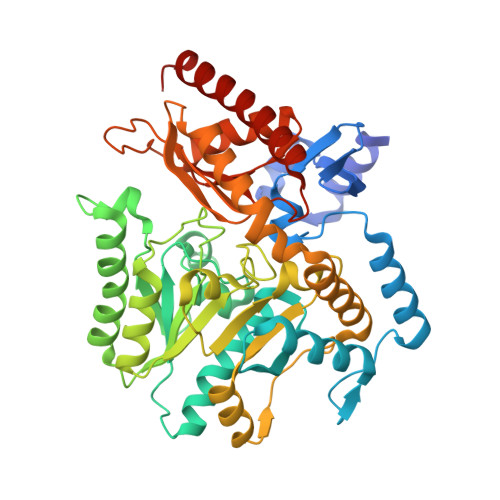
    6S4G

  • PubMed Abstract: 

    One of the main factors hampering the implementation in industry of transaminase-based processes for the synthesis of enantiopure amines is their often low storage and operational stability. Our still limited understanding of the inactivation processes undermining the stability of wild-type transaminases represents an obstacle to improving their stability through enzyme engineering. In this paper we present a model describing the inactivation process of the well-characterized (S)-selective amine transaminase from Chromobacterium violaceum. The cornerstone of the model, supported by structural, computational, mutagenesis and biophysical data, is the central role of the catalytic lysine as a conformational switch. Upon breakage of the lysine-PLP Schiff base, the strain associated with the catalytically active lysine conformation is dissipated in a slow relaxation process capable of triggering the known structural rearrangements occurring in the holo-to-apo transition and ultimately promoting dimer dissociation. Due to the occurrence in the literature of similar PLP-dependent inactivation models valid for other non-transaminase enzymes belonging to the same fold-class, the role of the catalytic lysine as conformational switch might extend beyond the transaminase enzyme group and offer new insight to drive future non-trivial engineering strategies.


  • Organizational Affiliation
    • Department of Industrial Biotechnology, KTH Royal Institute of Technology, AlbaNova University Center, SE-106 91, Stockholm, Sweden.

Macromolecule Content 

  • Total Structure Weight: 209.95 kDa 
  • Atom Count: 14,848 
  • Modeled Residue Count: 1,815 
  • Deposited Residue Count: 1,860 
  • Unique protein chains: 1

Macromolecules

Find similar proteins by:|  3D Structure
Entity ID: 1
MoleculeChains  Sequence LengthOrganismDetailsImage
Probable aminotransferase
A, B, C, D
465Chromobacterium violaceum ATCC 12472Mutation(s): 0 
Gene Names: CV_2025
EC: 2.6.1.62
UniProt
Find proteins for Q7NWG4 (Chromobacterium violaceum (strain ATCC 12472 / DSM 30191 / JCM 1249 / CCUG 213 / NBRC 12614 / NCIMB 9131 / NCTC 9757 / MK))
Explore Q7NWG4 
Go to UniProtKB:  Q7NWG4
Entity Groups
Sequence Clusters30% Identity50% Identity70% Identity90% Identity95% Identity100% Identity
UniProt GroupQ7NWG4
Sequence Annotations
Expand
Reference Sequence

Small Molecules

Ligands 3 Unique
IDChains Name / Formula / InChI Key2D Diagram3D Interactions
PMP
(Subject of Investigation/LOI)

Query on PMP


Download:Ideal Coordinates CCD File
E [auth A],
G [auth B],
J [auth C],
L [auth D]
4'-DEOXY-4'-AMINOPYRIDOXAL-5'-PHOSPHATE
C8 H13 N2 O5 P
ZMJGSOSNSPKHNH-UHFFFAOYSA-N
PEG

Query on PEG


Download:Ideal Coordinates CCD File
I [auth B],
K [auth C]
DI(HYDROXYETHYL)ETHER
C4 H10 O3
MTHSVFCYNBDYFN-UHFFFAOYSA-N
EDO

Query on EDO


Download:Ideal Coordinates CCD File
F [auth A],
H [auth B],
M [auth D],
N [auth D],
O [auth D]
1,2-ETHANEDIOL
C2 H6 O2
LYCAIKOWRPUZTN-UHFFFAOYSA-N

Experimental Data & Validation

Experimental Data

  • Method: X-RAY DIFFRACTION
  • Resolution: 1.67 Å
  • R-Value Free:  0.185 (Depositor), 0.195 (DCC) 
  • R-Value Work:  0.161 (Depositor), 0.173 (DCC) 
  • R-Value Observed: 0.162 (Depositor) 
Space Group: P 1
Unit Cell:
Length ( Å )Angle ( ˚ )
a = 60.909α = 75.07
b = 61.973β = 81.31
c = 118.81γ = 75.3
Software Package:
Software NamePurpose
REFMACrefinement
XDSdata reduction
Aimlessdata scaling
PHASERphasing
PDB_EXTRACTdata extraction

Structure Validation

View Full Validation Report


Ligand Structure Quality Assessment 


Entry History 

& Funding Information

Deposition Data


Funding OrganizationLocationGrant Number
European UnionSweden634200

Revision History  (Full details and data files)

  • Version 1.0: 2019-07-17
    Type: Initial release
  • Version 1.1: 2019-12-04
    Changes: Database references
  • Version 1.2: 2024-01-24
    Changes: Data collection, Database references, Refinement description