6RRR | pdb_00006rrr

Crystal structure of the tyrosinase PvdP from Pseudomonas aeruginosa


Experimental Data Snapshot

  • Method: X-RAY DIFFRACTION
  • Resolution: 2.11 Å
  • R-Value Free: 
    0.220 (Depositor), 0.228 (DCC) 
  • R-Value Work: 
    0.169 (Depositor), 0.178 (DCC) 
  • R-Value Observed: 
    0.172 (Depositor) 

wwPDB Validation 3D Report Full Report

Validation slider image for 6RRR

This is version 1.1 of the entry. See complete history

Literature

A novel mechanism of inhibition by phenylthiourea on PvdP, a tyrosinase synthesizing pyoverdine of Pseudomonas aeruginosa.

Wibowo, J.P.Batista, F.A.van Oosterwijk, N.Groves, M.R.Dekker, F.J.Quax, W.J.

(2020) Int J Biol Macromol 146: 212-221

  • DOI: https://doi.org/10.1016/j.ijbiomac.2019.12.252
  • Primary Citation Related Structures: 
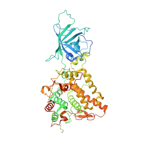
    6RRP, 6RRQ, 6RRR

  • PubMed Abstract: 

    The biosynthesis of pyoverdine, the major siderophore of Pseudomonas aeruginosa, is a well-organized process involving a discrete number of enzyme-catalyzed steps. The final step of this process involves the PvdP tyrosinase, which converts ferribactin into pyoverdine. Thus, inhibition of the PvdP tyrosinase activity provides an attractive strategy to interfere with siderophore synthesis to manage P. aeruginosa infections. Here, we report phenylthiourea as a non-competitive inhibitor of PvdP for which we solved a crystal structure in complex with PvdP. The crystal structure indicates that phenylthiourea binds to an allosteric binding site and thereby interferes with its tyrosinase activity. We further provide proofs that PvdP tyrosinase inhibition by phenylthiourea requires the C-terminal lid region. This provides opportunities to develop inhibitors that target the allosteric site, which seems to be confined to fluorescent pseudomonads, and not the tyrosinase active site. Furthermore, increases the chances to identify PvdP inhibitors that selectively interfere with siderophore synthesis.


  • Organizational Affiliation
    • Department of Chemical and Pharmaceutical Biology, Groningen Research Institute of Pharmacy, University of Groningen, the Netherlands; Faculty of Pharmacy, University of Muhammadiyah Banjarmasin, Banjarmasin, Indonesia.

Macromolecule Content 

  • Total Structure Weight: 125.1 kDa 
  • Atom Count: 8,161 
  • Modeled Residue Count: 932 
  • Deposited Residue Count: 1,088 
  • Unique protein chains: 1

Macromolecules

Find similar proteins by:|  3D Structure
Entity ID: 1
MoleculeChains  Sequence LengthOrganismDetailsImage
PvdP
A, B
544Pseudomonas aeruginosa PAO1Mutation(s): 0 
Gene Names: pvdPPA2392
UniProt
Find proteins for Q9I188 (Pseudomonas aeruginosa (strain ATCC 15692 / DSM 22644 / CIP 104116 / JCM 14847 / LMG 12228 / 1C / PRS 101 / PAO1))
Explore Q9I188 
Go to UniProtKB:  Q9I188
Entity Groups
Sequence Clusters30% Identity50% Identity70% Identity90% Identity95% Identity100% Identity
UniProt GroupQ9I188
Sequence Annotations
Expand
Reference Sequence

Small Molecules

Modified Residues  1 Unique
IDChains TypeFormula2D DiagramParent
CME
Query on CME
A, B
L-PEPTIDE LINKINGC5 H11 N O3 S2CYS

Experimental Data & Validation

Experimental Data

  • Method: X-RAY DIFFRACTION
  • Resolution: 2.11 Å
  • R-Value Free:  0.220 (Depositor), 0.228 (DCC) 
  • R-Value Work:  0.169 (Depositor), 0.178 (DCC) 
  • R-Value Observed: 0.172 (Depositor) 
Space Group: P 1 21 1
Unit Cell:
Length ( Å )Angle ( ˚ )
a = 44.162α = 90
b = 112.616β = 92.71
c = 100.265γ = 90
Software Package:
Software NamePurpose
REFMACrefinement
XDSdata reduction
Aimlessdata scaling
MOLREPphasing
PDB_EXTRACTdata extraction

Structure Validation

View Full Validation Report


Entry History 

Deposition Data

Revision History  (Full details and data files)

  • Version 1.0: 2020-04-15
    Type: Initial release
  • Version 1.1: 2024-10-09
    Changes: Data collection, Database references, Structure summary