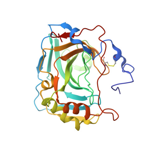
Structural comparison of protiated, H/D-exchanged and deuterated human carbonic anhydrase IX.
Koruza, K., Lafumat, B., Nyblom, M., Mahon, B.P., Knecht, W., McKenna, R., Fisher, S.Z.(2019) Acta Crystallogr D Struct Biol 75: 895-903
- PubMed: 31588921 Search on PubMedSearch on PubMed Central
- DOI: https://doi.org/10.1107/S2059798319010027
- Primary Citation Related Structures:
6RQN, 6RQQ, 6RQU, 6RQW - PubMed Abstract:
Human carbonic anhydrase IX (CA IX) expression is upregulated in hypoxic solid tumours, promoting cell survival and metastasis. This observation has made CA IX a target for the development of CA isoform-selective inhibitors. To enable structural studies of CA IX-inhibitor complexes using X-ray and neutron crystallography, a CA IX surface variant (CA IX SV ; the catalytic domain with six surface amino-acid substitutions) has been developed that can be routinely crystallized. Here, the preparation of protiated (H/H), H/D-exchanged (H/D) and deuterated (D/D) CA IX SV for crystallographic studies and their structural comparison are described. Four CA IX SV X-ray crystal structures are compared: two H/H crystal forms, an H/D crystal form and a D/D crystal form. The overall active-site organization in each version is essentially the same, with only minor positional changes in active-site solvent, which may be owing to deuteration and/or resolution differences. Analysis of the crystal contacts and packing reveals different arrangements of CA IX SV compared with previous reports. To our knowledge, this is the first report comparing three different deuterium-labelled crystal structures of the same protein, marking an important step in validating the active-site structure of CA IX SV for neutron protein crystallography.
- Department of Biology and Lund Protein Production Platform, Lund University, Sölvegatan 35, 223 62 Lund, Sweden.
Organizational Affiliation: