6RIV | pdb_00006riv

Crystal structure of Alopecurus myosuroides GSTF


Experimental Data Snapshot

  • Method: X-RAY DIFFRACTION
  • Resolution: 1.33 Å
  • R-Value Free: 
    0.163 (Depositor), 0.176 (DCC) 
  • R-Value Work: 
    0.131 (Depositor) 
  • R-Value Observed: 
    0.133 (Depositor) 

Starting Model: experimental
View more details

wwPDB Validation 3D Report Full Report

Validation slider image for 6RIV

Ligand Structure Quality Assessment 


This is version 1.2 of the entry. See complete history

Literature

Comparative structural and functional analysis of phi class glutathione transferases involved in multiple-herbicide resistance of grass weeds and crops.

Georgakis, N.Poudel, N.Papageorgiou, A.C.Labrou, N.E.

(2020) Plant Physiol Biochem 149: 266-276

  • DOI: https://doi.org/10.1016/j.plaphy.2020.02.012
  • Primary Citation Related Structures: 
    6RIV

  • PubMed Abstract: 

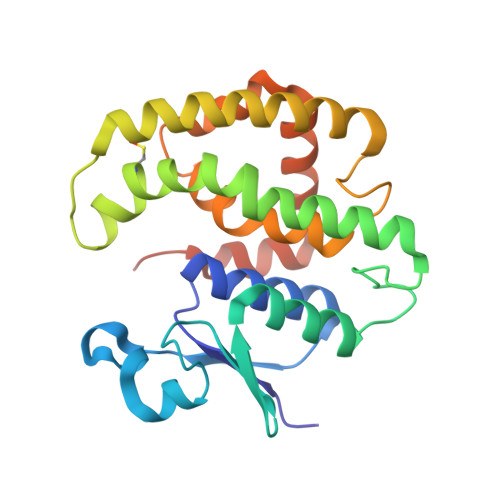
    Multiple-herbicide resistant (MHR) weeds are a global problem and a looming threat to weed control in crops. MHR weeds express a specific phi class glutathione transferase (MHR-GSTF) which seems to contribute to herbicide resistance. The present work aims to investigate the structure and catalytic properties of the MHR-GSTFs from different grass weeds and crops (Alopecurus myosuroides, Lolium rigidum, Hordeum vulgare, Triticum aestivum). Recombinant MHR-GSTFs were expressed in E. coli and purified by affinity chromatography. Kinetic analysis of substrate specificity using a range of thiol substrates and xenobiotic compounds suggested that all enzymes display a broad range of specificity and are capable of detoxifying major stress-induced toxic products. Notably, all tested enzymes exhibited high activity towards organic hydroperoxides. The crystal structure of MHR-GSTF from Alopecurus myosuroides (AmGSTF) was determined by molecular replacement at 1.33 Å resolution. The enzyme was resolved with bound glutathione sulfenic acid (GSOH) at the G-site and succinic acid at the H-site. The enzyme shows conserved structural features compared to other Phi class GSTs. However, some differences were observed at the C-terminal helix H9 that may affect substrate specificity. The structural and functional features of AmGSTF were compared with those of the homologue crop enzymes (HvGSTF and TaGSTF) and discussed in light of their contribution to the MHR mechanism.


  • Organizational Affiliation
    • Laboratory of Enzyme Technology, Department of Biotechnology, School of Applied Biology and Biotechnology, Agricultural University of Athens, 75 Iera Odos Street, GR-11855, Athens, Greece.

Macromolecule Content 

  • Total Structure Weight: 53.1 kDa 
  • Atom Count: 4,008 
  • Modeled Residue Count: 434 
  • Deposited Residue Count: 454 
  • Unique protein chains: 1

Macromolecules

Find similar proteins by:|  3D Structure
Entity ID: 1
MoleculeChains  Sequence LengthOrganismDetailsImage
Glutathione transferaseA [auth B],
B [auth A]
227Alopecurus myosuroidesMutation(s): 0 
Gene Names: GST2c
EC: 2.5.1.18
UniProt
Find proteins for Q9ZS17 (Alopecurus myosuroides)
Explore Q9ZS17 
Go to UniProtKB:  Q9ZS17
Entity Groups
Sequence Clusters30% Identity50% Identity70% Identity90% Identity95% Identity100% Identity
UniProt GroupQ9ZS17
Sequence Annotations
Expand
Reference Sequence

Small Molecules

Ligands 4 Unique
IDChains Name / Formula / InChI Key2D Diagram3D Interactions
GS8

Query on GS8


Download:Ideal Coordinates CCD File
C [auth B],
I [auth A]
S-Hydroxy-Glutathione
C10 H17 N3 O7 S
PFXSQLOWBQWLCX-WDSKDSINSA-N
SIN

Query on SIN


Download:Ideal Coordinates CCD File
D [auth B],
J [auth A]
SUCCINIC ACID
C4 H6 O4
KDYFGRWQOYBRFD-UHFFFAOYSA-N
GOL

Query on GOL


Download:Ideal Coordinates CCD File
E [auth B]GLYCEROL
C3 H8 O3
PEDCQBHIVMGVHV-UHFFFAOYSA-N
NA

Query on NA


Download:Ideal Coordinates CCD File
F [auth B]
G [auth B]
H [auth B]
K [auth A]
L [auth A]
F [auth B],
G [auth B],
H [auth B],
K [auth A],
L [auth A],
M [auth A]
SODIUM ION
Na
FKNQFGJONOIPTF-UHFFFAOYSA-N
Modified Residues  1 Unique
IDChains TypeFormula2D DiagramParent
CSO
Query on CSO
A [auth B],
B [auth A]
L-PEPTIDE LINKINGC3 H7 N O3 SCYS

Experimental Data & Validation

Experimental Data

  • Method: X-RAY DIFFRACTION
  • Resolution: 1.33 Å
  • R-Value Free:  0.163 (Depositor), 0.176 (DCC) 
  • R-Value Work:  0.131 (Depositor) 
  • R-Value Observed: 0.133 (Depositor) 
Space Group: P 1
Unit Cell:
Length ( Å )Angle ( ˚ )
a = 50.289α = 97.74
b = 50.708β = 110.24
c = 51.603γ = 110.96
Software Package:
Software NamePurpose
REFMACrefinement
PDB_EXTRACTdata extraction
XDSdata reduction
Aimlessdata scaling
MrBUMPphasing

Structure Validation

View Full Validation Report


Ligand Structure Quality Assessment 


Entry History 

Deposition Data

Revision History  (Full details and data files)

  • Version 1.0: 2020-03-04
    Type: Initial release
  • Version 1.1: 2024-01-24
    Changes: Data collection, Database references, Derived calculations, Refinement description
  • Version 1.2: 2024-10-16
    Changes: Structure summary