6RCI | pdb_00006rci

Crystal structure of REXO2


Experimental Data Snapshot

  • Method: X-RAY DIFFRACTION
  • Resolution: 2.00 Å
  • R-Value Free: 
    0.233 (Depositor), 0.239 (DCC) 
  • R-Value Work: 
    0.199 (Depositor), 0.205 (DCC) 
  • R-Value Observed: 
    0.200 (Depositor) 

wwPDB Validation 3D Report Full Report

Validation slider image for 6RCI

This is version 1.3 of the entry. See complete history

Literature

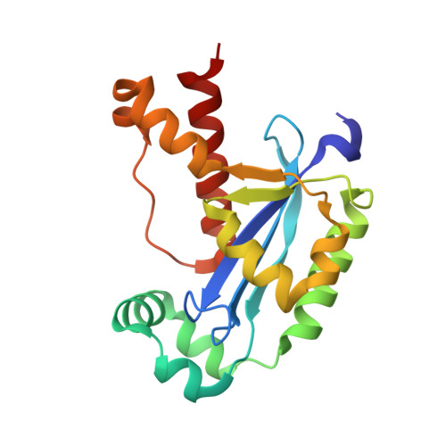
Dinucleotide Degradation by REXO2 Maintains Promoter Specificity in Mammalian Mitochondria.

Nicholls, T.J.Spahr, H.Jiang, S.Siira, S.J.Koolmeister, C.Sharma, S.Kauppila, J.H.K.Jiang, M.Kaever, V.Rackham, O.Chabes, A.Falkenberg, M.Filipovska, A.Larsson, N.G.Gustafsson, C.M.

(2019) Mol Cell 76: 784-796.e6

  • DOI: https://doi.org/10.1016/j.molcel.2019.09.010
  • Primary Citation Related Structures: 
    6RCI, 6RCL, 6RCN

  • PubMed Abstract: 

    Oligoribonucleases are conserved enzymes that degrade short RNA molecules of up to 5 nt in length and are assumed to constitute the final stage of RNA turnover. Here we demonstrate that REXO2 is a specialized dinucleotide-degrading enzyme that shows no preference between RNA and DNA dinucleotide substrates. A heart- and skeletal-muscle-specific knockout mouse displays elevated dinucleotide levels and alterations in gene expression patterns indicative of aberrant dinucleotide-primed transcription initiation. We find that dinucleotides act as potent stimulators of mitochondrial transcription initiation in vitro. Our data demonstrate that increased levels of dinucleotides can be used to initiate transcription, leading to an increase in transcription levels from both mitochondrial promoters and other, nonspecific sequence elements in mitochondrial DNA. Efficient RNA turnover by REXO2 is thus required to maintain promoter specificity and proper regulation of transcription in mammalian mitochondria.


  • Organizational Affiliation
    • Department of Medical Biochemistry and Cell Biology, University of Gothenburg, PO Box 440, Gothenburg 405 30, Sweden.

Macromolecule Content 

  • Total Structure Weight: 41.79 kDa 
  • Atom Count: 2,910 
  • Modeled Residue Count: 326 
  • Deposited Residue Count: 360 
  • Unique protein chains: 1

Macromolecules

Find similar proteins by:|  3D Structure
Entity ID: 1
MoleculeChains  Sequence LengthOrganismDetailsImage
Oligoribonuclease, mitochondrial
A, B
180Homo sapiensMutation(s): 0 
Gene Names: REXO2SFNSMFNCGI-114
EC: 3.1 (PDB Primary Data), 3.1.15 (UniProt)
UniProt & NIH Common Fund Data Resources
Find proteins for Q9Y3B8 (Homo sapiens)
Explore Q9Y3B8 
Go to UniProtKB:  Q9Y3B8
PHAROS:  Q9Y3B8
GTEx:  ENSG00000076043 
Entity Groups
Sequence Clusters30% Identity50% Identity70% Identity90% Identity95% Identity100% Identity
UniProt GroupQ9Y3B8
Sequence Annotations
Expand
Reference Sequence

Experimental Data & Validation

Experimental Data

  • Method: X-RAY DIFFRACTION
  • Resolution: 2.00 Å
  • R-Value Free:  0.233 (Depositor), 0.239 (DCC) 
  • R-Value Work:  0.199 (Depositor), 0.205 (DCC) 
  • R-Value Observed: 0.200 (Depositor) 
Space Group: C 2 2 21
Unit Cell:
Length ( Å )Angle ( ˚ )
a = 36.164α = 90
b = 128.492β = 90
c = 170.22γ = 90
Software Package:
Software NamePurpose
XSCALEdata scaling
BUSTERrefinement
PDB_EXTRACTdata extraction
XDSdata reduction
PHASERphasing

Structure Validation

View Full Validation Report


Entry History 

& Funding Information

Deposition Data


Funding OrganizationLocationGrant Number
Max Planck SocietyGermany--
Swedish Research CouncilSweden--
Knut and Alice Wallenberg FoundationSweden--
European Research Council--

Revision History  (Full details and data files)

  • Version 1.0: 2019-10-09
    Type: Initial release
  • Version 1.1: 2019-10-16
    Changes: Data collection, Database references
  • Version 1.2: 2019-12-18
    Changes: Database references
  • Version 1.3: 2024-05-15
    Changes: Data collection, Database references