6R2O | pdb_00006r2o

Hemoglobin structure from serial crystallography with a 3D-printed nozzle.


Experimental Data Snapshot

  • Method: X-RAY DIFFRACTION
  • Resolution: 2.46 Å
  • R-Value Free: 
    0.212 (Depositor), 0.214 (DCC) 
  • R-Value Work: 
    0.166 (Depositor), 0.168 (DCC) 
  • R-Value Observed: 
    0.170 (Depositor) 

Starting Model: experimental
View more details

wwPDB Validation 3D Report Full Report

Validation slider image for 6R2O

Ligand Structure Quality Assessment 


This is version 1.3 of the entry. See complete history

Literature

Ultracompact 3D microfluidics for time-resolved structural biology.

Knoska, J.Adriano, L.Awel, S.Beyerlein, K.R.Yefanov, O.Oberthuer, D.Pena Murillo, G.E.Roth, N.Sarrou, I.Villanueva-Perez, P.Wiedorn, M.O.Wilde, F.Bajt, S.Chapman, H.N.Heymann, M.

(2020) Nat Commun 11: 657-657

  • DOI: https://doi.org/10.1038/s41467-020-14434-6
  • Primary Citation Related Structures: 
    6R2O

  • PubMed Abstract: 

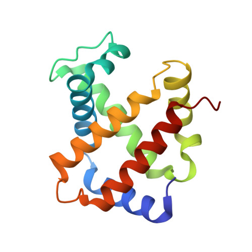
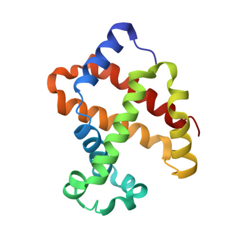
    To advance microfluidic integration, we present the use of two-photon additive manufacturing to fold 2D channel layouts into compact free-form 3D fluidic circuits with nanometer precision. We demonstrate this technique by tailoring microfluidic nozzles and mixers for time-resolved structural biology at X-ray free-electron lasers (XFELs). We achieve submicron jets with speeds exceeding 160 m s -1 , which allows for the use of megahertz XFEL repetition rates. By integrating an additional orifice, we implement a low consumption flow-focusing nozzle, which is validated by solving a hemoglobin structure. Also, aberration-free in operando X-ray microtomography is introduced to study efficient equivolumetric millisecond mixing in channels with 3D features integrated into the nozzle. Such devices can be printed in minutes by locally adjusting print resolution during fabrication. This technology has the potential to permit ultracompact devices and performance improvements through 3D flow optimization in all fields of microfluidic engineering.


  • Organizational Affiliation
    • CFEL, Center for Free-Electron Laser Science, Deutsches Elektronen-Synchrotron DESY, Notkestrasse 85, 22607, Hamburg, Germany.

Macromolecule Content 

  • Total Structure Weight: 64.72 kDa 
  • Atom Count: 4,797 
  • Modeled Residue Count: 572 
  • Deposited Residue Count: 572 
  • Unique protein chains: 2

Macromolecules

Find similar proteins by:|  3D Structure
Entity ID: 1
MoleculeChains  Sequence LengthOrganismDetailsImage
Hemoglobin subunit alpha
A, C
141Equus caballusMutation(s): 0 
UniProt
Find proteins for P01958 (Equus caballus)
Explore P01958 
Go to UniProtKB:  P01958
Entity Groups
Sequence Clusters30% Identity50% Identity70% Identity90% Identity95% Identity100% Identity
UniProt GroupP01958
Sequence Annotations
Expand
Reference Sequence
Find similar proteins by:|  3D Structure
Entity ID: 2
MoleculeChains  Sequence LengthOrganismDetailsImage
Hemoglobin subunit beta
B, D
145Equus caballusMutation(s): 0 
UniProt
Find proteins for P02062 (Equus caballus)
Explore P02062 
Go to UniProtKB:  P02062
Entity Groups
Sequence Clusters30% Identity50% Identity70% Identity90% Identity95% Identity100% Identity
UniProt GroupP02062
Sequence Annotations
Expand
Reference Sequence

Small Molecules

Ligands 4 Unique
IDChains Name / Formula / InChI Key2D Diagram3D Interactions
HEM

Query on HEM


Download:Ideal Coordinates CCD File
E [auth A],
G [auth B],
H [auth C],
K [auth D]
PROTOPORPHYRIN IX CONTAINING FE
C34 H32 Fe N4 O4
KABFMIBPWCXCRK-RGGAHWMASA-L
PEG

Query on PEG


Download:Ideal Coordinates CCD File
L [auth D]DI(HYDROXYETHYL)ETHER
C4 H10 O3
MTHSVFCYNBDYFN-UHFFFAOYSA-N
CL

Query on CL


Download:Ideal Coordinates CCD File
J [auth C]CHLORIDE ION
Cl
VEXZGXHMUGYJMC-UHFFFAOYSA-M
NA

Query on NA


Download:Ideal Coordinates CCD File
F [auth A],
I [auth C]
SODIUM ION
Na
FKNQFGJONOIPTF-UHFFFAOYSA-N

Experimental Data & Validation

Experimental Data

  • Method: X-RAY DIFFRACTION
  • Resolution: 2.46 Å
  • R-Value Free:  0.212 (Depositor), 0.214 (DCC) 
  • R-Value Work:  0.166 (Depositor), 0.168 (DCC) 
  • R-Value Observed: 0.170 (Depositor) 
Space Group: P 21 21 21
Unit Cell:
Length ( Å )Angle ( ˚ )
a = 62.5α = 90
b = 80.8β = 90
c = 110.8γ = 90
Software Package:
Software NamePurpose
PHENIXrefinement
CrystFELdata reduction
CrystFELdata scaling
PHASERphasing

Structure Validation

View Full Validation Report


Ligand Structure Quality Assessment 


Entry History 

Deposition Data

Revision History  (Full details and data files)

  • Version 1.0: 2020-01-29
    Type: Initial release
  • Version 1.1: 2020-02-19
    Changes: Database references
  • Version 1.2: 2023-12-13
    Changes: Data collection, Database references, Derived calculations, Refinement description
  • Version 1.3: 2024-01-24
    Changes: Refinement description