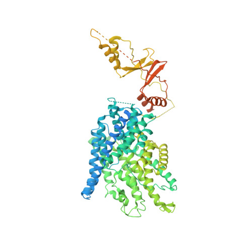
Structure of the human ClC-1 chloride channel.
Wang, K., Preisler, S.S., Zhang, L., Cui, Y., Missel, J.W., Gronberg, C., Gotfryd, K., Lindahl, E., Andersson, M., Calloe, K., Egea, P.F., Klaerke, D.A., Pusch, M., Pedersen, P.A., Zhou, Z.H., Gourdon, P.(2019) PLoS Biol 17: e3000218-e3000218
- PubMed: 31022181
- DOI: https://doi.org/10.1371/journal.pbio.3000218
- Primary Citation of Related Structures:
6QV6, 6QVB, 6QVC, 6QVD, 6QVU - PubMed Abstract:
ClC-1 protein channels facilitate rapid passage of chloride ions across cellular membranes, thereby orchestrating skeletal muscle excitability. Malfunction of ClC-1 is associated with myotonia congenita, a disease impairing muscle relaxation. Here, we present the cryo-electron microscopy (cryo-EM) structure of human ClC-1, uncovering an architecture reminiscent of that of bovine ClC-K and CLC transporters. The chloride conducting pathway exhibits distinct features, including a central glutamate residue ("fast gate") known to confer voltage-dependence (a mechanistic feature not present in ClC-K), linked to a somewhat rearranged central tyrosine and a narrower aperture of the pore toward the extracellular vestibule. These characteristics agree with the lower chloride flux of ClC-1 compared with ClC-K and enable us to propose a model for chloride passage in voltage-dependent CLC channels. Comparison of structures derived from protein studied in different experimental conditions supports the notion that pH and adenine nucleotides regulate ClC-1 through interactions between the so-called cystathionine-β-synthase (CBS) domains and the intracellular vestibule ("slow gating"). The structure also provides a framework for analysis of mutations causing myotonia congenita and reveals a striking correlation between mutated residues and the phenotypic effect on voltage gating, opening avenues for rational design of therapies against ClC-1-related diseases.
- Department of Biomedical Sciences, University of Copenhagen, Copenhagen, Denmark.
Organizational Affiliation:


