6QS1 | pdb_00006qs1

Crystal structure of human Angiotensin-1 converting enzyme N-domain in complex with BPPb


Experimental Data Snapshot

  • Method: X-RAY DIFFRACTION
  • Resolution: 1.80 Å
  • R-Value Free: 
    0.232 (Depositor), 0.234 (DCC) 
  • R-Value Work: 
    0.202 (Depositor), 0.203 (DCC) 
  • R-Value Observed: 
    0.202 (Depositor) 

Starting Model: experimental
View more details

wwPDB Validation 3D Report Full Report

Validation slider image for 6QS1

Ligand Structure Quality Assessment 


This is version 3.2 of the entry. See complete history

Literature

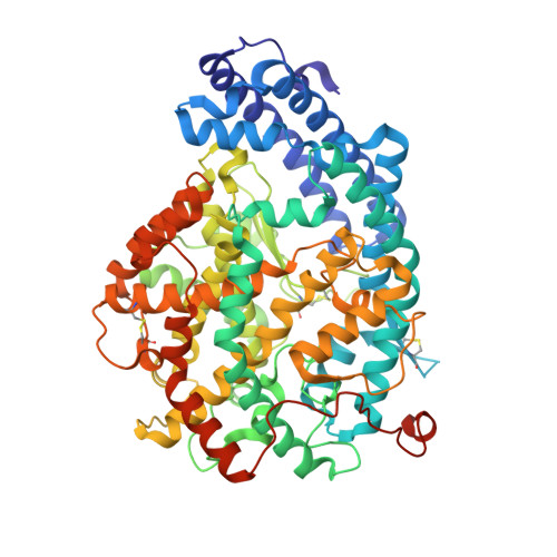
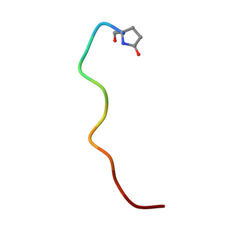
Structural basis for the C-domain-selective angiotensin-converting enzyme inhibition by bradykinin-potentiating peptide b (BPPb).

Sturrock, E.D.Lubbe, L.Cozier, G.E.Schwager, S.L.U.Arowolo, A.T.Arendse, L.B.Belcher, E.Acharya, K.R.

(2019) Biochem J 476: 1553-1570

  • DOI: https://doi.org/10.1042/BCJ20190290
  • Primary Citation Related Structures: 
    6QS1

  • PubMed Abstract: 

    Angiotensin-converting enzyme (ACE) is a zinc metalloprotease best known for its role in blood pressure regulation. ACE consists of two homologous catalytic domains, the N- and C-domain, that display distinct but overlapping catalytic functions in vivo owing to subtle differences in substrate specificity. While current generation ACE inhibitors target both ACE domains, domain-selective ACE inhibitors may be clinically advantageous, either reducing side effects or having utility in new indications. Here, we used site-directed mutagenesis, an ACE chimera and X-ray crystallography to unveil the molecular basis for C-domain-selective ACE inhibition by the bradykinin-potentiating peptide b (BPPb), naturally present in Brazilian pit viper venom. We present the BPPb N-domain structure in comparison with the previously reported BPPb C-domain structure and highlight key differences in peptide interactions with the S 4 to S 9 subsites. This suggests the involvement of these subsites in conferring C-domain-selective BPPb binding, in agreement with the mutagenesis results where unique residues governing differences in active site exposure, lid structure and dynamics between the two domains were the major drivers for C-domain-selective BPPb binding. Mere disruption of BPPb interactions with unique S 2 and S 4 subsite residues, which synergistically assist in BPPb binding, was insufficient to abolish C-domain selectivity. The combination of unique S 9 -S 4 and S 2 ' subsite C-domain residues was required for the favourable entry, orientation and thus, selective binding of the peptide. This emphasizes the need to consider factors other than direct protein-inhibitor interactions to guide the design of domain-selective ACE inhibitors, especially in the case of larger peptides.


  • Organizational Affiliation
    • Department of Integrative Biomedical Sciences, Institute of Infectious Disease and Molecular Medicine, University of Cape Town, Observatory, 7925, Cape Town, Republic of South Africa edward.sturrock@uct.ac.za.

Macromolecule Content 

  • Total Structure Weight: 152.63 kDa 
  • Atom Count: 11,129 
  • Modeled Residue Count: 1,227 
  • Deposited Residue Count: 1,280 
  • Unique protein chains: 2

Macromolecules

Find similar proteins by:|  3D Structure
Entity ID: 1
MoleculeChains  Sequence LengthOrganismDetailsImage
Angiotensin-converting enzyme
A, B
629Homo sapiensMutation(s): 8 
Gene Names: ACEDCPDCP1
EC: 3.2.1 (PDB Primary Data), 3.4.15.1 (PDB Primary Data)
UniProt & NIH Common Fund Data Resources
Find proteins for P12821 (Homo sapiens)
Explore P12821 
Go to UniProtKB:  P12821
PHAROS:  P12821
GTEx:  ENSG00000159640 
Entity Groups
Sequence Clusters30% Identity50% Identity70% Identity90% Identity95% Identity100% Identity
UniProt GroupP12821
Glycosylation
Glycosylation Sites: 3Go to GlyGen: P12821-1
Sequence Annotations
Expand
Reference Sequence
Find similar proteins by:  Sequence   |   3D Structure  
Entity ID: 2
MoleculeChains  Sequence LengthOrganismDetailsImage
Bradykinin potentiating peptide bC [auth E],
D [auth F]
11Gloydius blomhoffiiMutation(s): 0 

Oligosaccharides

Help  
Entity ID: 3
MoleculeChains Length2D Diagram GlycosylationD Interactions
2-acetamido-2-deoxy-beta-D-glucopyranose-(1-4)-2-acetamido-2-deoxy-beta-D-glucopyranoseE [auth C]2N-Glycosylation
Glycosylation Resources
GlyTouCan: G42666HT
GlyCosmos: G42666HT
GlyGen: G42666HT
Entity ID: 4
MoleculeChains Length2D Diagram GlycosylationD Interactions
beta-D-mannopyranose-(1-4)-2-acetamido-2-deoxy-beta-D-glucopyranose-(1-4)-[alpha-L-fucopyranose-(1-6)]2-acetamido-2-deoxy-beta-D-glucopyranoseF [auth D]4N-Glycosylation
Glycosylation Resources
GlyTouCan: G32152BH
GlyCosmos: G32152BH
GlyGen: G32152BH
Entity ID: 5
MoleculeChains Length2D Diagram GlycosylationD Interactions
2-acetamido-2-deoxy-beta-D-glucopyranose-(1-4)-[alpha-L-fucopyranose-(1-6)]2-acetamido-2-deoxy-beta-D-glucopyranose
G, H
3N-Glycosylation
Glycosylation Resources
GlyTouCan: G21290RB
GlyCosmos: G21290RB
GlyGen: G21290RB
Entity ID: 6
MoleculeChains Length2D Diagram GlycosylationD Interactions
alpha-L-fucopyranose-(1-6)-2-acetamido-2-deoxy-beta-D-glucopyranose
I
2N-Glycosylation
Glycosylation Resources
GlyTouCan: G86851RC
GlyCosmos: G86851RC
GlyGen: G86851RC

Small Molecules

Ligands 9 Unique
IDChains Name / Formula / InChI Key2D Diagram3D Interactions
XPE

Query on XPE


Download:Ideal Coordinates CCD File
J [auth A]3,6,9,12,15,18,21,24,27-NONAOXANONACOSANE-1,29-DIOL
C20 H42 O11
DTPCFIHYWYONMD-UHFFFAOYSA-N
NAG

Query on NAG


Download:Ideal Coordinates CCD File
Z [auth B]2-acetamido-2-deoxy-beta-D-glucopyranose
C8 H15 N O6
OVRNDRQMDRJTHS-FMDGEEDCSA-N
PGE

Query on PGE


Download:Ideal Coordinates CCD File
T [auth A]TRIETHYLENE GLYCOL
C6 H14 O4
ZIBGPFATKBEMQZ-UHFFFAOYSA-N
PEG

Query on PEG


Download:Ideal Coordinates CCD File
BA [auth B],
CA [auth B],
M [auth A],
Q [auth A]
DI(HYDROXYETHYL)ETHER
C4 H10 O3
MTHSVFCYNBDYFN-UHFFFAOYSA-N
ZN

Query on ZN


Download:Ideal Coordinates CCD File
HA [auth B],
V [auth A]
ZINC ION
Zn
PTFCDOFLOPIGGS-UHFFFAOYSA-N
EDO

Query on EDO


Download:Ideal Coordinates CCD File
AA [auth B]
GA [auth B]
K [auth A]
L [auth A]
R [auth A]
AA [auth B],
GA [auth B],
K [auth A],
L [auth A],
R [auth A],
S [auth A]
1,2-ETHANEDIOL
C2 H6 O2
LYCAIKOWRPUZTN-UHFFFAOYSA-N
BO3

Query on BO3


Download:Ideal Coordinates CCD File
DA [auth B]
EA [auth B]
FA [auth B]
N [auth A]
O [auth A]
DA [auth B],
EA [auth B],
FA [auth B],
N [auth A],
O [auth A],
P [auth A],
U [auth A]
BORIC ACID
B H3 O3
KGBXLFKZBHKPEV-UHFFFAOYSA-N
CL

Query on CL


Download:Ideal Coordinates CCD File
IA [auth B],
JA [auth B],
W [auth A],
X [auth A]
CHLORIDE ION
Cl
VEXZGXHMUGYJMC-UHFFFAOYSA-M
MG

Query on MG


Download:Ideal Coordinates CCD File
KA [auth B],
Y [auth A]
MAGNESIUM ION
Mg
JLVVSXFLKOJNIY-UHFFFAOYSA-N
Modified Residues  1 Unique
IDChains TypeFormula2D DiagramParent
PCA
Query on PCA
C [auth E],
D [auth F]
L-PEPTIDE LINKINGC5 H7 N O3GLN

Experimental Data & Validation

Experimental Data

  • Method: X-RAY DIFFRACTION
  • Resolution: 1.80 Å
  • R-Value Free:  0.232 (Depositor), 0.234 (DCC) 
  • R-Value Work:  0.202 (Depositor), 0.203 (DCC) 
  • R-Value Observed: 0.202 (Depositor) 
Space Group: P 1
Unit Cell:
Length ( Å )Angle ( ˚ )
a = 72.682α = 88.86
b = 77.07β = 64.59
c = 82.363γ = 75.03
Software Package:
Software NamePurpose
PHENIXrefinement
DIALSdata reduction
Aimlessdata scaling
PHASERphasing

Structure Validation

View Full Validation Report


Ligand Structure Quality Assessment 


Entry History 

& Funding Information

Deposition Data


Funding OrganizationLocationGrant Number
Medical Research Council (United Kingdom)United KingdomMR/M026647/1

Revision History  (Full details and data files)

  • Version 1.0: 2019-05-22
    Type: Initial release
  • Version 1.1: 2019-06-12
    Changes: Data collection, Database references
  • Version 1.2: 2019-08-21
    Changes: Data collection
  • Version 2.0: 2020-03-11
    Changes: Data collection, Derived calculations, Polymer sequence
  • Version 3.0: 2020-07-29
    Type: Remediation
    Reason: Carbohydrate remediation
    Changes: Atomic model, Data collection, Derived calculations, Structure summary
  • Version 3.1: 2024-01-24
    Changes: Data collection, Database references, Refinement description, Structure summary
  • Version 3.2: 2024-11-13
    Changes: Structure summary