6QPQ | pdb_00006qpq

The structure of the cohesin head module elucidates the mechanism of ring opening


Experimental Data Snapshot

  • Method: X-RAY DIFFRACTION
  • Resolution: 2.10 Å
  • R-Value Free: 
    0.251 (DCC) 
  • R-Value Work: 
    0.218 (DCC) 

wwPDB Validation 3D Report Full Report

Validation slider image for 6QPQ

This is version 1.3 of the entry. See complete history

Literature

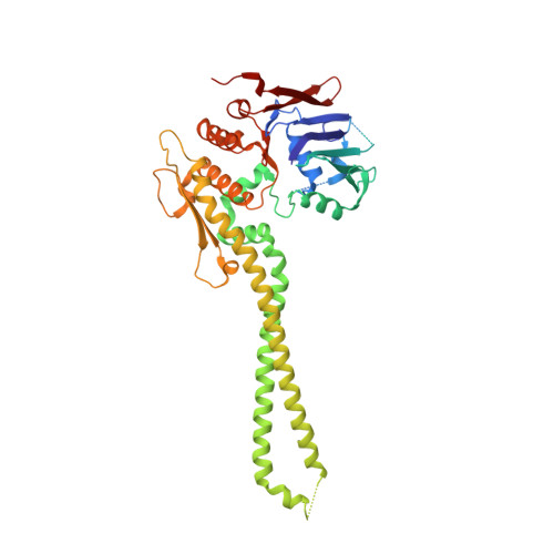
The structure of the cohesin ATPase elucidates the mechanism of SMC-kleisin ring opening.

Muir, K.W.Li, Y.Weis, F.Panne, D.

(2020) Nat Struct Mol Biol 27: 233-239

  • DOI: https://doi.org/10.1038/s41594-020-0379-7
  • Primary Citation Related Structures: 
    6QPQ, 6QPW

  • PubMed Abstract: 

    Genome regulation requires control of chromosome organization by SMC-kleisin complexes. The cohesin complex contains the Smc1 and Smc3 subunits that associate with the kleisin Scc1 to form a ring-shaped complex that can topologically engage chromatin to regulate chromatin structure. Release from chromatin involves opening of the ring at the Smc3-Scc1 interface in a reaction that is controlled by acetylation and engagement of the Smc ATPase head domains. To understand the underlying molecular mechanisms, we have determined the 3.2-Å resolution cryo-electron microscopy structure of the ATPγS-bound, heterotrimeric cohesin ATPase head module and the 2.1-Å resolution crystal structure of a nucleotide-free Smc1-Scc1 subcomplex from Saccharomyces cerevisiae and Chaetomium thermophilium. We found that ATP-binding and Smc1-Smc3 heterodimerization promote conformational changes within the ATPase that are transmitted to the Smc coiled-coil domains. Remodeling of the coiled-coil domain of Smc3 abrogates the binding surface for Scc1, thus leading to ring opening at the Smc3-Scc1 interface.


  • Organizational Affiliation
    • European Molecular Biology Laboratory, Grenoble, France. kmuir@mrc-lmb.cam.ac.uk.

Macromolecule Content 

  • Total Structure Weight: 230.47 kDa 
  • Atom Count: 8,097 
  • Modeled Residue Count: 957 
  • Deposited Residue Count: 2,038 
  • Unique protein chains: 2

Macromolecules

Find similar proteins by:|  3D Structure
Entity ID: 1
MoleculeChains  Sequence LengthOrganismDetailsImage
Structural maintenance of chromosomes protein,Structural maintenance of chromosomes protein
A, C
453Thermochaetoides thermophila DSM 1495Mutation(s): 0 
Gene Names: CTHT_0066330
UniProt
Find proteins for G0SGH3 (Chaetomium thermophilum (strain DSM 1495 / CBS 144.50 / IMI 039719))
Explore G0SGH3 
Go to UniProtKB:  G0SGH3
Entity Groups
Sequence Clusters30% Identity50% Identity70% Identity90% Identity95% Identity100% Identity
UniProt GroupG0SGH3
Sequence Annotations
Expand
Reference Sequence
Find similar proteins by:|  3D Structure
Entity ID: 2
MoleculeChains  Sequence LengthOrganismDetailsImage
Sister chromatid cohesion protein 1
B, D
566Saccharomyces cerevisiae S288CMutation(s): 0 
Gene Names: MCD1PDS3RHC21SCC1YDL003WYD8119.04
UniProt
Find proteins for Q12158 (Saccharomyces cerevisiae (strain ATCC 204508 / S288c))
Explore Q12158 
Go to UniProtKB:  Q12158
Entity Groups
Sequence Clusters30% Identity50% Identity70% Identity90% Identity95% Identity100% Identity
UniProt GroupQ12158
Sequence Annotations
Expand
Reference Sequence

Experimental Data & Validation

Experimental Data

  • Method: X-RAY DIFFRACTION
  • Resolution: 2.10 Å
  • R-Value Free:  0.251 (DCC) 
  • R-Value Work:  0.218 (DCC) 
Space Group: P 2 21 21
Unit Cell:
Length ( Å )Angle ( ˚ )
a = 80.752α = 90
b = 111.13β = 90
c = 166.195γ = 90
Software Package:
Software NamePurpose
PHENIXrefinement
XDSdata reduction
Aimlessdata scaling
PHASERphasing

Structure Validation

View Full Validation Report


Entry History 

Deposition Data

Revision History  (Full details and data files)

  • Version 1.0: 2020-02-05
    Type: Initial release
  • Version 1.1: 2020-02-26
    Changes: Database references
  • Version 1.2: 2020-03-18
    Changes: Database references
  • Version 1.3: 2024-05-15
    Changes: Data collection, Database references