6QMN | pdb_00006qmn

Crystal structure of a Ribonuclease A-Onconase chimera


Experimental Data Snapshot

  • Method: X-RAY DIFFRACTION
  • Resolution: 2.31 Å
  • R-Value Free: 
    0.294 (Depositor), 0.303 (DCC) 
  • R-Value Work: 
    0.205 (Depositor), 0.214 (DCC) 
  • R-Value Observed: 
    0.209 (Depositor) 

Starting Model: experimental
View more details

wwPDB Validation 3D Report Full Report

Validation slider image for 6QMN

This is version 1.3 of the entry. See complete history

Literature

Structure, stability and aggregation propensity of a Ribonuclease A-Onconase chimera.

Esposito, L.Donnarumma, F.Ruggiero, A.Leone, S.Vitagliano, L.Picone, D.

(2019) Int J Biol Macromol 133: 1125-1133

  • DOI: https://doi.org/10.1016/j.ijbiomac.2019.04.164
  • Primary Citation Related Structures: 
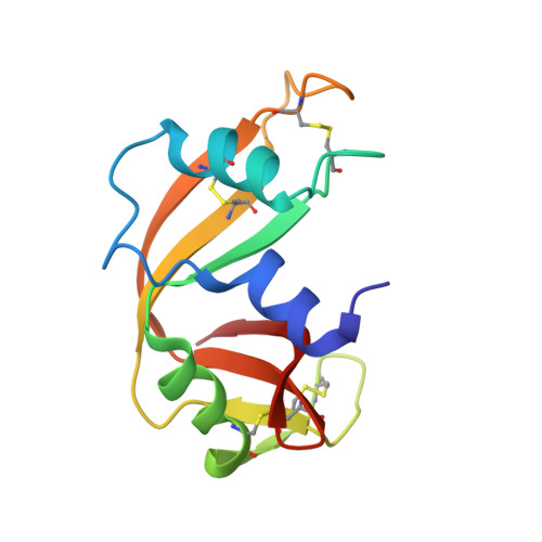
    6QMN

  • PubMed Abstract: 

    Structural roles of loop regions are frequently overlooked in proteins. Nevertheless, they may be key players in the definition of protein topology and in the self-assembly processes occurring through domain swapping. We here investigate the effects on structure and stability of replacing the loop connecting the last two β-strands of RNase A with the corresponding region of the more thermostable Onconase. The crystal structure of this chimeric variant (RNaseA-ONC) shows that its terminal loop size better adheres to the topological rules for the design of stabilized proteins, proposed by Baker and coworkers [43]. Indeed, RNaseA-ONC displays a thermal stability close to that of RNase A, despite the lack of Pro at position 114, which, due to its propensity to favor a cis peptide bond, has been identified as an important stabilizing factor of the native protein. Accordingly, RNaseA-ONC is significantly more stable than RNase A variants lacking Pro114; RNaseA-ONC also displays a higher propensity to form oligomers in native conditions when compared to either RNase A or Onconase. This finding demonstrates that modifications of terminal loops should to be carefully controlled in terms of size and sequence to avoid unwanted and/or potentially harmful aggregation processes.


  • Organizational Affiliation
    • CNR Istituto di Biostrutture e Bioimmagini, Via Mezzocannone 16, I-80134 Napoli, Italy. Electronic address: luciana.esposito@cnr.it.

Macromolecule Content 

  • Total Structure Weight: 40.76 kDa 
  • Atom Count: 3,003 
  • Modeled Residue Count: 366 
  • Deposited Residue Count: 366 
  • Unique protein chains: 1

Macromolecules

Find similar proteins by:|  3D Structure
Entity ID: 1
MoleculeChains  Sequence LengthOrganismDetailsImage
Ribonuclease pancreatic
A, B, C
122Bison bisonMutation(s): 0 
Gene Names: RNASE1RNS1
EC: 3.1.27.5 (PDB Primary Data), 4.6.1.18 (UniProt)
UniProt
Find proteins for P61824 (Bison bison)
Explore P61824 
Go to UniProtKB:  P61824
Entity Groups
Sequence Clusters30% Identity50% Identity70% Identity90% Identity95% Identity100% Identity
UniProt GroupP61824
Sequence Annotations
Expand
Reference Sequence

Experimental Data & Validation

Experimental Data

  • Method: X-RAY DIFFRACTION
  • Resolution: 2.31 Å
  • R-Value Free:  0.294 (Depositor), 0.303 (DCC) 
  • R-Value Work:  0.205 (Depositor), 0.214 (DCC) 
  • R-Value Observed: 0.209 (Depositor) 
Space Group: P 1 21 1
Unit Cell:
Length ( Å )Angle ( ˚ )
a = 58.442α = 90
b = 51.796β = 93.01
c = 66.093γ = 90
Software Package:
Software NamePurpose
REFMACrefinement
PDB_EXTRACTdata extraction
HKL-2000data reduction
HKL-2000data scaling
PHASERphasing

Structure Validation

View Full Validation Report


Entry History 

Deposition Data

Revision History  (Full details and data files)

  • Version 1.0: 2019-05-08
    Type: Initial release
  • Version 1.1: 2019-05-22
    Changes: Data collection, Database references
  • Version 1.2: 2024-01-24
    Changes: Data collection, Database references, Refinement description
  • Version 1.3: 2024-10-16
    Changes: Structure summary