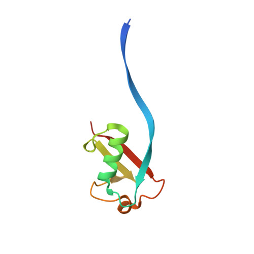
6QK9

A dimeric ubiquitin formed by a single amino acid substitution


Experimental Data Snapshot

  • Method: X-RAY DIFFRACTION
  • Resolution: 2.23 Å
  • R-Value Free: 0.281 
  • R-Value Work: 0.239 
  • R-Value Observed: 0.241 

Starting Model: experimental
View more details

wwPDB Validation   3D Report Full Report


This is version 1.2 of the entry. See complete history


Literature

Identification and Characterization of Mutations in Ubiquitin Required for Non-covalent Dimer Formation.

Gabrielsen, M.Buetow, L.Kowalczyk, D.Zhang, W.Sidhu, S.S.Huang, D.T.

(2019) Structure 27: 1452-1459.e4

  • DOI: https://doi.org/10.1016/j.str.2019.06.008
  • Primary Citation of Related Structures:  
    6QK9

  • PubMed Abstract: 

    Ubiquitin (Ub) is a small protein that post-translationally modifies a variety of substrates in eukaryotic cells to modulate substrate function. The ability of Ub to interact with numerous protein domains makes Ub an attractive scaffold for engineering ubiquitin variants (UbVs) with high target specificity. Previously, we identified a UbV that formed a non-covalent stable dimer via a β-strand exchange, and in the current work we identified and characterized the minimal substitutions in the primary sequence of Ub required to form a higher ordered complex. Using solution angle scattering and X-ray crystallography, we show that a single substitution of residue Gly10 to either Ala or Val is sufficient to convert Ub from a monomer to a dimer. We also investigate contributions to dimer formation by the residues in the surrounding sequence. These results can be used to develop next-generation phage-display libraries of UbVs to engineer new interfaces for protein recognition.


  • Organizational Affiliation

    Cancer Research UK Beatson Institute, Institute of Cancer Sciences, University of Glasgow, Garscube Estate, Switchback Road, Glasgow G61 1BD, UK.


Macromolecules
Find similar proteins by:  (by identity cutoff)  |  3D Structure
Entity ID: 1
MoleculeChains Sequence LengthOrganismDetailsImage
Polyubiquitin-B
A, B, C, D, E
A, B, C, D, E, F, G, H, I, J, K, L
79Homo sapiensMutation(s): 1 
Gene Names: UBB
UniProt & NIH Common Fund Data Resources
Find proteins for P0CG47 (Homo sapiens)
Explore P0CG47 
Go to UniProtKB:  P0CG47
PHAROS:  P0CG47
GTEx:  ENSG00000170315 
Entity Groups  
Sequence Clusters30% Identity50% Identity70% Identity90% Identity95% Identity100% Identity
UniProt GroupP0CG47
Sequence Annotations
Expand
  • Reference Sequence
Experimental Data & Validation

Experimental Data

  • Method: X-RAY DIFFRACTION
  • Resolution: 2.23 Å
  • R-Value Free: 0.281 
  • R-Value Work: 0.239 
  • R-Value Observed: 0.241 
  • Space Group: P 21 21 21
Unit Cell:
Length ( Å )Angle ( ˚ )
a = 83.934α = 90
b = 87.262β = 90
c = 109.841γ = 90
Software Package:
Software NamePurpose
PHENIXrefinement
xia2data reduction
Aimlessdata scaling
MoRDaphasing

Structure Validation

View Full Validation Report


Entry History & Funding Information

Deposition Data


Funding OrganizationLocationGrant Number
Cancer Research UKUnited KingdomA23278
European Research CouncilUnited Kingdom647849

Revision History  (Full details and data files)

  • Version 1.0: 2019-07-24
    Type: Initial release
  • Version 1.1: 2019-09-18
    Changes: Data collection, Database references
  • Version 1.2: 2024-01-24
    Changes: Data collection, Database references, Refinement description