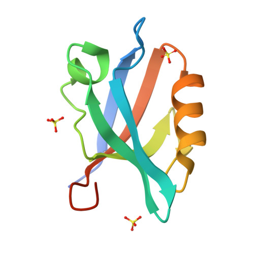
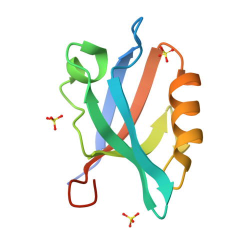
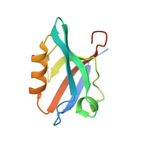
6QJJ

Crystal Structure of the third PDZ domain of PSD-95 protein: space group P3221


Experimental Data Snapshot

  • Method: X-RAY DIFFRACTION
  • Resolution: 1.70 Å
  • R-Value Free: 0.223 
  • R-Value Work: 0.202 
  • R-Value Observed: 0.203 

Starting Model: experimental
View more details

wwPDB Validation   3D Report Full Report


This is version 1.3 of the entry. See complete history

Re-refinement Note

This entry reflects an alternative modeling of the original data in: 5OIG


Literature

Conformational changes in the third PDZ domain of the neuronal postsynaptic density protein 95.

Camara-Artigas, A.Murciano-Calles, J.Martinez, J.C.

(2019) Acta Crystallogr D Struct Biol 75: 381-391

  • DOI: https://doi.org/10.1107/S2059798319001980
  • Primary Citation of Related Structures:  
    6QJD, 6QJF, 6QJG, 6QJI, 6QJJ, 6QJK, 6QJL, 6QJN

  • PubMed Abstract: 

    PDZ domains are protein-protein recognition modules that interact with other proteins through short sequences at the carboxyl terminus. These domains are structurally characterized by a conserved fold composed of six β-strands and two α-helices. The third PDZ domain of the neuronal postsynaptic density protein 95 has an additional α-helix (α3), the role of which is not well known. In previous structures, a succinimide was identified in the β2-β3 loop instead of Asp332. The presence of this modified residue results in conformational changes in α3. In this work, crystallographic structures of the following have been solved: a truncated form of the third PDZ domain of the neuronal postsynaptic density protein 95 from which α3 has been removed, D332P and D332G variants of the protein, and a new crystal form of this domain showing the binding of Asp332 to the carboxylate-binding site of a symmetry-related molecule. Crystals of the wild type and variants were obtained in different space groups, which reflects the conformational plasticity of the domain. Indeed, the overall analysis of these structures suggests that the conformation of the β2-β3 loop is correlated with the fold acquired by α3. The alternate conformation of the β2-β3 loop affects the electrostatics of the carboxylate-binding site and might modulate the binding of different PDZ-binding motifs.


  • Organizational Affiliation

    Department of Chemistry and Physics, Agrifood Campus of International Excellence (ceiA3) and CIAMBITAL, University of Almería, Carretera de Sacramento, 04120 Almeria, Spain.


Macromolecules
Find similar proteins by:  (by identity cutoff)  |  3D Structure
Entity ID: 1
MoleculeChains Sequence LengthOrganismDetailsImage
Disks large homolog 499Homo sapiensMutation(s): 0 
Gene Names: DLG4PSD95
UniProt & NIH Common Fund Data Resources
Find proteins for P78352 (Homo sapiens)
Explore P78352 
Go to UniProtKB:  P78352
PHAROS:  P78352
GTEx:  ENSG00000132535 
Entity Groups  
Sequence Clusters30% Identity50% Identity70% Identity90% Identity95% Identity100% Identity
UniProt GroupP78352
Sequence Annotations
Expand
  • Reference Sequence
Experimental Data & Validation

Experimental Data

  • Method: X-RAY DIFFRACTION
  • Resolution: 1.70 Å
  • R-Value Free: 0.223 
  • R-Value Work: 0.202 
  • R-Value Observed: 0.203 
  • Space Group: P 32 2 1
Unit Cell:
Length ( Å )Angle ( ˚ )
a = 34.076α = 90
b = 34.076β = 90
c = 113.672γ = 120
Software Package:
Software NamePurpose
XDSdata reduction
Aimlessdata scaling
PHASERphasing
PHENIXrefinement
PDB_EXTRACTdata extraction
Cootmodel building

Structure Validation

View Full Validation Report


Entry History & Funding Information

Deposition Data


Funding OrganizationLocationGrant Number
Spanish Ministry of Economy and CompetitivenessSpainBIO2016-78020-R

Revision History  (Full details and data files)

  • Version 1.0: 2019-04-17
    Type: Initial release
  • Version 1.1: 2019-04-24
    Changes: Data collection, Database references
  • Version 1.2: 2020-01-15
    Changes: Data collection
  • Version 1.3: 2024-01-24
    Changes: Data collection, Database references, Refinement description